
J. Mater. Sci. Technol. ›› 2021, Vol. 63: 54-61.DOI: 10.1016/j.jmst.2020.02.059
• Research Article • Previous Articles Next Articles
Erica Rosellaa,b, Nan Jiaa, Diego Mantovanib,c, Jesse Greenera,c,*( )
)
Received:2019-12-05
Revised:2020-01-27
Accepted:2020-02-09
Published:2021-02-10
Online:2021-02-15
Contact:
Jesse Greener
About author:*Laboratory, Department of Chemistry, Laval University, Québec, QC, Canada.E-mail address: Jesse.Greener@chm.ulaval.ca (J. Greener).Erica Rosella, Nan Jia, Diego Mantovani, Jesse Greener. A microfluidic approach for development of hybrid collagen-chitosan extracellular matrix-like membranes for on-chip cell cultures[J]. J. Mater. Sci. Technol., 2021, 63: 54-61.
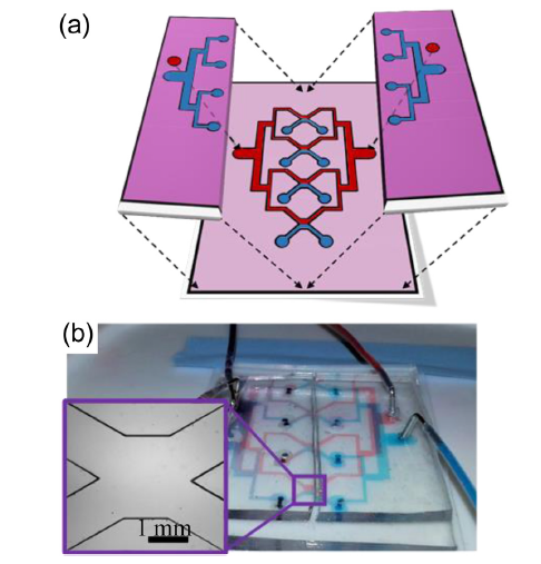
Fig. 1. Microfluidic platform for synthesis of biopolymer membranes within standing pH gradients. (a) Schematic of a parallel membrane synthesis device with 4 X-channels showing co-flowing basic solutions (blue) and the acidic biopolymer solution (red). (b) Photo of the realized device with dye solutions injected to visualize the co-flow patterns described in (a). Inset image shows a single X-channel before membrane synthesis.

Fig. 2. Viscosity measurements for (a) collagen solution in acid acetic 0.02 N at 4 (▲), 2 (?) and 1 mg mL-1 (●) and (b) chitosan at 5 (▲), 2 (?), and 1 mg mL-1 (●) concentrations. The vertical axis is the same scale as in (a). Both collagen and chitosan solutions were maintained at pH 3.5.
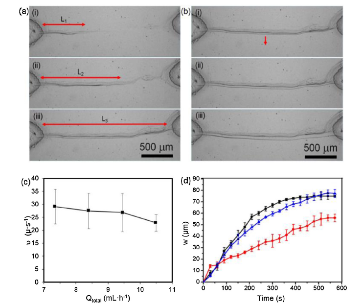
Fig. 3. In situ formation of chitosan membranes studied by time-lapse imaging. (a) Sequential images from the time-lapse series showing propagation of a chitosan membrane (growth parallel to flow indicated by red arrow) during the initial contact of the chitosan solution (5 mL h-1, pH 3.5) with NaOH solution (pH 10). Flow rates were Qp = 0.4 mL h-1 and Qb = 7.8 mL h-1 (Qtotal = 8.2 mL h-1). Red arrows show the position of the propagation front after each 15 s interval. (b) Average propagation velocity of a chitosan membrane measured for different solution total flow rates (Qtotal) while maintaining a flow rate ratio of Qb/Qp = 20. (c) Widening chitosan membrane in time (growth perpendicular to flow indicated by red arrow). The direction of flow is from left to right in images (a) and (c). (d) Membrane width versus time for 4 mL h-1 collagen (black) at Qb = 1.83 mL h-1, 5 mL h-1 chitosan (blue) at Qb = 2.7 mL h-1, and 60:40 (v/v%) collagen/chitosan hybrid (red) at Qb = 2.1 mL h-1. All pH values were 3.5 and 10 for the polymer and NaOH solutions, respectively. Error bars in (b) and (d) were generated from the standard deviation of quadruplicate measurements under identical conditions.
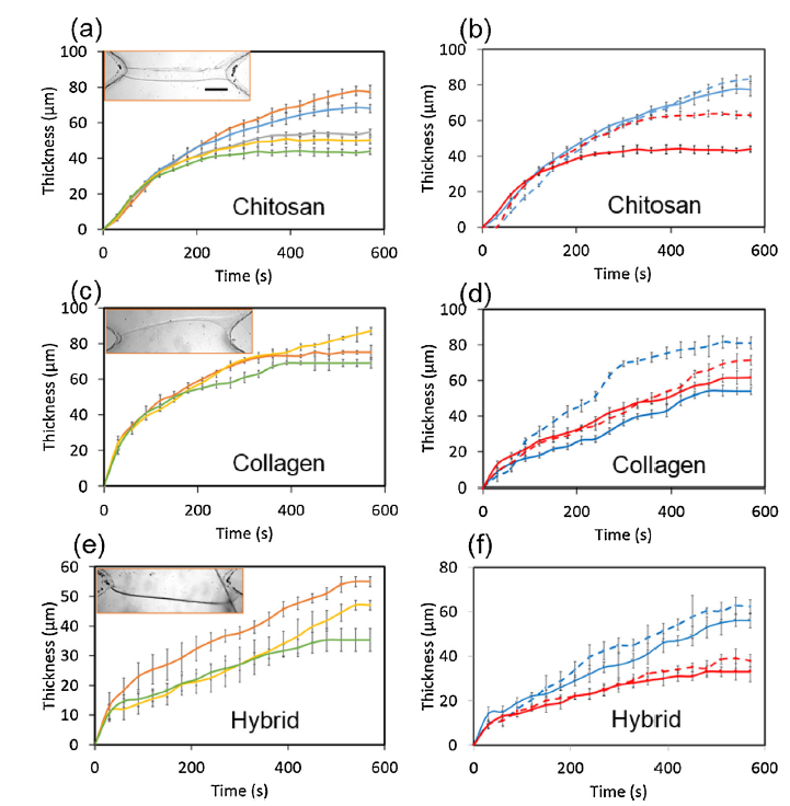
Fig. 4. Growth of membrane width and changes to uniformity under different flow rates for chitosan (a, b), collagen (c, d) and collagen/chitosan hybrids (e, f) during the first 10 min of growth. Inset images showing typical membranes after 30 min. (scale bar = 500 μm). (a) Chitosan membrane formation was tested under five different flow rates: Qtotal = 2.7 (orange), 5.4 (blue), 8.1 (gray), 10.8 (yellow), and 13.5 mL h-1 (green). (b) Width measurements of chitosan membranes at the upstream (solid line) and downstream (dashed line) positions in the X-channel for fastest (red) and slowest (blue) flow rates used in (a). (c) Collagen membrane formation was tested under three different flow rates: Qtotal = 0.63 (orange), 1.23 (yellow), and 1.83 mL h-1 (green). (d) Width measurements of collagen at upstream and downstream positions, with line colours same as in (b). (e) Hybrid chitosan-collagen membrane formation for three different solution flow rates: Qtotal = 1.03 (orange), 2.1 (yellow), and 5 mL h-1 (green). (f) Width measurements of hybrid chitosan-collagen membrane at upstream and downstream positions, with line colours same as in (b). All flow rate ratios were Qb/Qp = 20.
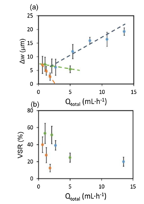
Fig. 5. (a) Variation in membrane width (Δw) between up- and downstream positions as a function of total flow rate. (b) Volume swelling ratios for membranes formed at different flow rates for 0.4 mL mg-1 collagen (orange circles), 0.4 mL mg-1 chitosan (blue circles), and 60/40 collagen/chitosan hybrid (green circles). Dashed lines in (a) are included for eye guidance.
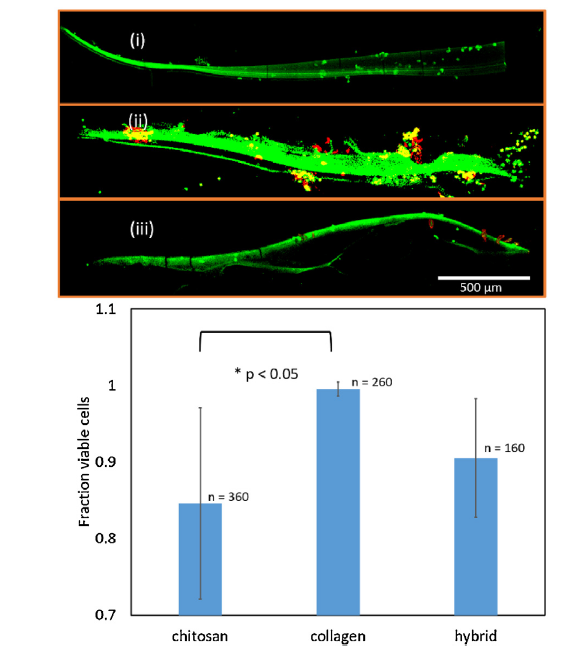
Fig. 6. (a) Live/dead (green/red) assays of 3T3 fibroblast cells one week after inoculation for (i) collagen, (ii) chitosan, and (iii) collagen/chitosan hybrid membranes. Yellow regions correspond to dead cells overlapping with a green background and are therefore, considered as red for the purposes of statistics. Membranes appear green due to retention of live stain and auto-fluorescence. Membrane synthesis was conducted with Qtotal = 0.63, 2.7, and 1.0 mL h-1 for collagen, chitosan, and collagen-chitosan hybrid extracellular membranes, respectively. Scale bar in (i) is common for (i-iii). (b) Cell viability calculated based on 4 repeated experiments for each membrane type. ANOVA analysis followed by post-hoc paired t-test for the same variance showed a significant difference between chitosan and collagen (*).
| [1] |
H.J. Kim, D. Huh, G. Hamilton, D.E. Ingber, Lab Chip 12 (2012) 2165-2174.
DOI URL |
| [2] |
D. Huh, B.D. Matthews, A. Mammoto, M. Montoya-Zavala, Y.H. Hong, D.E. Ingber, Science 328 (2010) 1662-1668.
DOI URL PMID |
| [3] |
F. Berthiaume, T.J. Maguire, M.L. Yarmush, Annu. Rev. Chem. Biomol. Eng. 2 (2011) 403-430.
DOI URL PMID |
| [4] | P. Fratzl, USA, 2018, pp. 1-12. |
| [5] |
A. Matteo, D. Mantovani, Polymers 2 (2010) 664-680.
DOI URL |
| [6] |
F. Boccafoschi, M. Bosetti, C. Mosca, D. Mantovani, M. Cannas, J. Tissue Eng. Regen. Med. 6 (2011) 60-67.
DOI URL PMID |
| [7] | S.T. Beyer, A. Bsoul, A. Ahmadi, K. Walus, 3D alginate constructs for tissue engineering printed using a coaxial flow focusing microfluidic device, Transducers & Eurosensors XXVII (2013) 1206-1209. |
| [8] |
R.N. Chen, G.M. Wang, C.H. Chen, H.O. Ho, M.T. Sheu, Biomacromolecules 7 (2006) 1058-1064.
DOI URL PMID |
| [9] |
A. Sionkowska, M. Wisniewski, J. Skopinska, C.J. Kennedy, T.J. Wess, Biomaterials 25 (2004) 795-801.
DOI URL |
| [10] |
Z. Chen, X. Mo, C. He, H. Wang, Carbohydr. Polym. 72 (2008) 410-418.
DOI URL |
| [11] |
Z. Chen, X. Mo, F. Qing, Material Letters. 61 (2007) 3490-3494.
DOI URL |
| [12] |
E. Kang, S.-J. Shin, K.H. Lee, S.-H. Lee, Lab Chip 10 (2010) 1856-1861.
DOI URL PMID |
| [13] |
S. Lin, W. Wang, X.-J. Ju, R. Xie, L.-Y. Chu, Lab Chip 14 (2014) 2626-2634.
DOI URL |
| [14] |
L. Li, Y. Nie, X. Shi, H. Wu, D. Ye, H. Chen, Biomicrofluidics 5 (2011) 036503-036508.
DOI URL |
| [15] | T. Cubaud, S. Notaro, Phys. Fluids 26 (2014) 1-10. |
| [16] |
X. Luo, D.L. Berlin, J. Betz, G.F. Payne, W.E. Bentley, G.W. Rubloff, Lab Chip 10 (2010) 59-65.
DOI URL PMID |
| [17] |
X. Luo, H.C. Wu, C.Y. Tsao, Y. Cheng, J. Betz, G.F. Payne, G.W. Rubloff, W.E. Bentley, Biomaterials 33 (2012) 5136-5143.
DOI URL |
| [18] |
N. Hakimi, R. Cheng, L. Leng, M. Sotoudehfar, P.Q. Ba, N. Bakhtyar, S. Amini-Nik, M.G. Jeschke, A. Günther, Lab Chip 18 (2018) 1440-1451.
DOI URL PMID |
| [19] |
Y.-M. Sun, W. Wang, Y.-Y. Wei, N.-N. Deng, Z. Liu, X.-J. Ju, R. Xie, L.-Y. Chu, Lab Chip 14 (2014) 2418-2427.
DOI URL |
| [20] |
M.J. Mondrinos, Y.-S. Yi, N.-K. Wu, X. Dinga, D. Huh, Lab Chip 17 (2017) 3146-3158.
DOI URL PMID |
| [21] |
F. Boccafoschi, N. Rajan, J. Habermehl, D. Mantovani, Macromol. Biosci. 7 (2007) 719-726.
DOI URL PMID |
| [22] |
D.I. Zeugolis, R.G. Paul, G. Attenburrow, J. Biomater. Sci. Polym. Ed. 20 (2009) 219-234.
DOI URL PMID |
| [23] |
B.A. Roeder, K. Kokini, J.E. Sturgis, J.P. Robinson, S.L. Voytik-Harbin, J. Biomech. Eng. 124 (2002) 214-222.
DOI URL PMID |
| [24] |
C. Sendner, R.R. Netz, Europhys. Lett. 81 (2008) 54006.
DOI URL |
| [25] |
C.E. Sing, A. Alexander-Katz, Europhys. Lett. 95 (2011) 48001.
DOI URL |
| [26] |
G.L. He, R. Messina, H.L. Löwen, A. Kiriy, V. Bocharova, M. Stamm, Soft Matter 5 (2009) 3014-3017.
DOI URL |
| [27] |
A. Lamura, R.G. Winkler, J. Chem. Phys. 137 (2012) 244909.
DOI URL PMID |
| [28] |
M. Harasim, B. Wunderlich, O. Peleg, M. Kröger, A.R. Bausch, Phys. Rev. Lett. 110 (2013) 108302.
DOI URL PMID |
| [29] |
L. Zhang, C. Ma, J. Sun, B. Shao, G. Protale, D. Chen, K. Liu, A. Herrmann, Angew. Chem. Int. Ed 57 (2018) 6878-6882.
DOI URL |
| [30] |
N. Jia, et al., A Microfluidic Approach to Micromembrane Synthesis for Complex Release Profiles of Nanocarriers, Lab on a Chip 20 (6) (2020) 1066-1071.
DOI URL PMID |
| [1] | Pengfei Ji, Bohan Chen, Bo Li, Yihao Tang, Guofeng Zhang, Xinyu Zhang, Mingzhen Ma, Riping Liu. Influence of Nb addition on microstructural evolution and compression mechanical properties of Ti-Zr alloys [J]. J. Mater. Sci. Technol., 2021, 69(0): 7-14. |
| [2] | Gopinathan Janarthanan, Insup Noh. Recent trends in metal ion based hydrogel biomaterials for tissue engineering and other biomedical applications [J]. J. Mater. Sci. Technol., 2021, 63(0): 35-53. |
| [3] | Dan He, Haiyan Li. Biomaterials affect cell-cell interactions in vitro in tissue engineering [J]. J. Mater. Sci. Technol., 2021, 63(0): 62-72. |
| [4] | Xian-Zong Wang, Hong-Qiang Fan, Triratna Muneshwar, Ken Cadien, Jing-Li Luo. Balancing the corrosion resistance and through-plane electrical conductivity of Cr coating via oxygen plasma treatment [J]. J. Mater. Sci. Technol., 2021, 61(0): 75-84. |
| [5] | Seung Woo Lee, Bongho Lee, Chaekyung Baik, Tae-Yang Kim, Chanho Pak. Multifunctional Ir-Ru alloy catalysts for reversal-tolerant anodes of polymer electrolyte membrane fuel cells [J]. J. Mater. Sci. Technol., 2021, 60(0): 105-112. |
| [6] | Jiahui Chen, Dainan Zhang, Song He, Gengpei Xia, Xiaoyi Wang, Quanjun Xiang, Tianlong Wen, Zhiyong Zhong, Yulong Liao. Thermal insulation design for efficient and scalable solar water interfacial evaporation and purification [J]. J. Mater. Sci. Technol., 2021, 66(0): 157-162. |
| [7] | Tao Liu, Caizhen Zhu, Wei Wu, Kai-Ning Liao, Xianjing Gong, Qijun Sun, Robert K.Y. Li. Facilely prepared layer-by-layer graphene membrane-based pressure sensor with high sensitivity and stability for smart wearable devices [J]. J. Mater. Sci. Technol., 2020, 45(0): 241-247. |
| [8] | Chengzhi Dai, Min Zhou, Weinan Jiang, Ximian Xiao, Jingcheng Zou, Yuxin Qian, Zihao Cong, Zhemin Ji, Longqiang Liu, Jiayang Xie, Zhongqian Qiao, Runhui Liu. Breaking or following the membrane-targeting mechanism: Exploring the antibacterial mechanism of host defense peptide mimicking poly(2-oxazoline)s [J]. J. Mater. Sci. Technol., 2020, 59(0): 220-226. |
| [9] | Xiao Lin, Yanjie Bai, Huan Zhou, Lei Yang. Mechano-active biomaterials for tissue repair and regeneration [J]. J. Mater. Sci. Technol., 2020, 59(0): 227-233. |
| [10] | Ruoxian Wang, Gaowu Qin, Erlin Zhang. Effect of Cu on Martensite Transformation of CoCrMo alloy for biomedical application [J]. J. Mater. Sci. Technol., 2020, 52(0): 127-135. |
| [11] | Hao Yu, Yi He, Guoqing Xiao, Yi Fan, Jing Ma, Yixuan Gao, Ruitong Hou, Jingyu Chen. Weak-reduction graphene oxide membrane for improving water purification performance [J]. J. Mater. Sci. Technol., 2020, 39(0): 106-112. |
| [12] | Jiuchuan Guo, Fanyu Zeng, Jinhong Guo, Xing Ma. Preparation and application of microfluidic SERS substrate: Challenges and future perspectives [J]. J. Mater. Sci. Technol., 2020, 37(0): 96-103. |
| [13] | Junlei Li, Ling Qin, Ke Yang, Zhijie Ma, Yongxuan Wang, Liangliang Cheng, Dewei Zhao. Materials evolution of bone plates for internal fixation of bone fractures: A review [J]. J. Mater. Sci. Technol., 2020, 36(0): 190-208. |
| [14] | Chang-Yang Li, Xiao-Li Fan, Rong-Chang Zeng, Lan-Yue Cui, Shuo-Qi Li, Fen Zhang, Qing-Kun He, M. Bobby Kannan, , Dong-Chu Chen, Shao-Kang Guan. Corrosion resistance of in-situ growth of nano-sized Mg(OH)2 on micro-arc oxidized magnesium alloy AZ31—Influence of EDTA [J]. J. Mater. Sci. Technol., 2019, 35(6): 1088-1098. |
| [15] | H.F. Li, F.L. Nie, Y.F. Zheng, Y. Cheng, S.C. Wei, R.Z. Valiev. Nanocrystalline Ti49.2Ni50.8 shape memory alloy as orthopaedic implant material with better performance [J]. J. Mater. Sci. Technol., 2019, 35(10): 2156-2162. |
| Viewed | ||||||
|
Full text |
|
|||||
|
Abstract |
|
|||||
 WeChat
