a School of Information and Communication Engineering, University of Electronic Science and Technology of China, Chengdu 610054, China
b Flexible Printed Electronic Technology Center & School of Materials Science and Engineering, Harbin Institute of Technology (Shenzhen), Shenzhen 518055, China
c Shenzhen Bay Laboratory, No. 9 Duxue Road, Shenzhen 518055, China
Lateral flow immunoassays (LFIAs) have been developed rapidly in recent years and used in a wide range of application at point-of-care-testing (POCT), where small biomolecules can be conveniently examined on a test strip. Compared with other biochemical detection methods such as ELISA (enzyme linked immunosorbent assay) or mass spectrometry method, LFIAs have the advantages of low cost, easy operation and short time-consuming. However, it suffers from low sensitivity since conventional LFIA can only realize qualitative detection based on colorimetric signals. With the increasing demand for more accurate and sensitive determination, novel nanomaterials have been used as labels in LFIAs due to their unique advantages in physical and chemical properties. Colloidal gold, fluorescent nanoparticles, SERS-active nanomaterials, magnetic nanoparticles and carbon nanomaterials are utilized in LFIAs to produce different kinds of signals for quantitative or semi-quantitative detection. This review paper first gives a description of the LFIA principles, and then focuses on the state-of-the-art nanomaterial labelling technology in LFIAs. At last, the conclusion and outlook are given to inspire exploration of more advanced nanomaterials for the development of future LFIAs.
Jiuchuan Guo, Shuqin Chen, Jinhong Guo, Xing Ma. Nanomaterial Labels in Lateral Flow Immunoassays for Point-of-Care-Testing. Journal of Materials Science & Technology[J], 2021, 60(0): 90-104 DOI:10.1016/j.jmst.2020.06.003
1. Introduction
The lateral flow immunoassay (LFIA) was emerged as a low-cost, rapid and simple detection method at the end of 1960s, and it has been used for analyzing small biomolecules at point-of-care-testing (POCT). The basic principle of it is to attach different labels to biomarkers (antigens or antibodies) to achieve macroscopic visualization or detection of microscopic immune responses by means of signal amplification. In 1976, the first radioimmunoassay based test strip was developed for the detection of human chorionic gonadotropin (hCG) in urine, and was introduced into the market in the United States. However, it wasn’t affordable for most of the average families at that time. In 1990s, in place of radioactive labels, Osikowicz et al. [1] used colored colloidal selenium for one-step chromatographic immunoassay determination of hCG in urine, which to a large extent reduce the fabrication cost. Since then, varieties of novel test strips have flooded into medical market for rapid immunoassay. Compared with traditional biochemical detection methods, LFIAs require no professional operation, much less detection time and flexible using conditions. However, it conventionally suffered from poor sensitivity and reproducibility, because the intensity of the produced signal is relatively low. Besides, since the colorimetric signals cannot be quantitatively determined by naked eyes, it was hard for LFIAs to achieve quantitative detection [2]. Hence, a growing concern has focused on the novel materials for labelling to realize signal amplification and quantification in LFIAs.
Nanomaterials, as an emerging type of material featured with size ranging from 1 to 100 nm, are considered to be ideal labels in bioanalysis and biosensing because of their unique physical and chemical properties. On the one hand, due to their much larger specific surface area than normal macroscopic particles, nanoparticles (NPs) can provide a large amount of space for surface modification, [3] and various molecular moieties can be attached to control their physical and chemical properties. On the other hand, NPs have unique advantages in optical, magnetic, electrical or thermal properties, which enable them to act as signal producers when they are combined with different analytes. The introduction of NP labels simplifies the inspection process of biomolecules and makes LFIAs more sensitive and selective. Currently, most commonly used nanomaterial in LFIAs is colloidal gold. Qualitative analysis can be achieved by observing the strip color with naked eyes. Although the method is simple and fast, it is difficult in accurate quantification [4,5]. In recent years, much effort has been made to develop new kinds of nanoparticle labels in order to make LFIAs more sensitive and accurate, and to be more adaptable to the today’s POCT. We have summarized the parameters of each nanoparticles mentioned in this paper in terms of their practical use in LFIAs, which is shown in Table 1. This review paper will briefly give a description of the structures of the LFIA test strips and illustrate the principles of two main detecting formats. And then, we will comprehensively discuss the state-of-the-art nanomaterial labels used in LFIA based on different detecting signals. At last, the conclusion and outlook will be highlighted. Compared with the previous reviews in this field [[73], [74], [75]], this paper generated a more comprehensive review of the current advanced nanomaterial labeling technology in LFIAs. Furthermore, we emphasized the importance and huge application prospects of multiplex detection in future’s LFIAs.
Table 1
Table 1Physical parameters comparisons among each nanoparticles
LFIA is a rapid diagnostic technology developed from enzyme 1inked immunosorbent assay (ELISA). It has the advantages of simple operation, low cost and short detecting time. The test strip of LFIA is combined with the sample pad, conjugate pad, nitrocellulose (NC) membrane and absorption pad (shown in Fig. 1). These are the places where immune reactions occur. We summarize the functions of each component as follows (shown in Table 2).
The general principle of LFIA is based on antibody-antigen specific interaction [5]. Specific materials are initially labelled on the antibodies. The labelled antibodies are then dispensed on the conjugate pad and will flow forward through capillary action when the sample liquid is applied on the sample pad. The liquid will pass through the test areas of the strip and finally reach to the adsorption pad at the end. Test line (TL) and control line (CL) are drawn over the NC membrane where the test result is displayed. We observe the test line to analyze the results and use control line to decide if the test is valid. In LFIAs, two formats are most commonly used for detecting: i) sandwich format, ii) competitive format.
2.1. Sandwich format LFIAs
In sandwich format LFIAs, three different antibodies are utilized. The primary antibody (Ab1) and secondary antibody (Ab2) are immobilized on the test line and the control line, respectively. When the sample liquid contains the target antigens (Ag), the Ag specifically bind to the labeled antibodies (Ab) on the conjugate pad to form an Ab-Ag complex. The Ab-Ag complex flow forwards on the NC membrane and will be captured by the Ab1 on the TL to form a sandwich format Ab-Ag-Ab1. As the Ab is labeled, we can examine the signals appearing on the TL to analyze the test result. As the liquid continues to move forward, the Ab2 immobilized on the CL which is known as a species-specific anti-immunoglobulin antibody, can catch the Ab to form the Ab-Ab2 complex. So, if the test is valid, signals always appear on the CL whether or not there are antigens contained in sample.
It is easy to understand that the sandwich format of LFIA requires at least two antigenic sites for the antigen, and thus, it is only suitable for large analytes.
2.2. Competitive format LFIA
Competitive format is used for testing small analytes which have only one antigenic site. Generally, this format utilizes two different antibodies and the format has two arrangements. For the first arrangement, the labeled antibody (Ab) is immobilized on the conjugate pad and the species-specific anti-immunoglobulin antibody (Ab2) is immobilized on the CL. Unlike the sandwich format, the TL in the competitive format is coated with the target antigen. When there are antigens in the sample liquid, the labeled Ab specifically bind to the antigens and flows through the NC membrane. Due to competitive inhibition, the antigens on the TL will not catch the flowing Ab-Ag complex, and thus signals on the TL decreases as the antibody concentration increases. In the case of no antigens contained in the sample, the labeled Ab is captured by the Ag on the TL, where strong signals will be produced. For the second arrangement, Ab is immobilized on the TL to capture the labeled Ag on the conjugated pad. When the sample liquid applied contains the unlabeled Ag, signals on the TL will greatly reduce to show a positive result because the unlabeled Ag bind onto the TL. Similar to the sandwich format, the CL is used for the validation of the test because of the formation of Ab-Ab2.
3. Nanomaterial labels in LFIA
For NP labeled LFIAs, it is critical to combine NPs and biomolecules in a stable manner. In the past few years, many studies have paved the way for the effective bonding process. Molina-Bolivar et al. [6] compared two approaches for immobilizing IgG antibodies on latex particles: physical adsorption and covalent binding. The results indicated that the covalent immunolatex had better immunoreactivity and stabilization. Gersten et al. [7] achieved the directly coupling of antibodies by the mediation of protein A, and MasanoriYoshioka et al. [8] introduced the antibody coupling mediated by glutaraldehyde served as a bifunctional coupling agent. However, these methods are easy to cause agglomeration of particles during the preparation process, and thus affecting test results. Currently, the most common and mature coupling method is EDC/NHS activation method, which is proposed by Steve Howell et al [9]. They used N-hydroxysuccinimide (NHS) and N'-(3-dimethylaminopropyl) carbodiimide hydrochloride (EDC) to activate the carboxyl group of the particles, which made it possible to link the amino group of the protein without introducing excess carbon chain. A large number of experiments have proved that this method has good stability and is easy to operate.
Based on the above achievements, various kinds of nanomaterials have been used in LFIAs for sensitive detecting, such as colloidal gold, fluorescent NPs, Raman probes, magnetic NPs and carbon NPs.
3.1. Colloidal gold
Colloidal gold refers to a gold nanoparticle suspension with a particle diameter between 1 to 150 nm. The gold nanoparticles (AuNPs) are commonly prepared by reducing the chloroauric acid (HAuCl4) solution. Different reduction methods determine the particle size, which leads to different color exhibition. AuNPs are easy to conjugate with immunoglobulins, toxins, antibiotics, hormones and other biological macromolecules through electrostatic interaction without affecting their biological activity.
Colloidal gold LFIAs (CG-LFIAs) emerged in the early 1980s, and the detection method is mainly based on color observation. Owing to the advantages of easy synthesis, rapid detection and low cost, CG-LFIAs have been widely used in POCT such as medical diagnosis [[10], [11], [12], [13], [14]], toxic detection [[15], [16], [17], [18]], food safety [[19], [20], [21], [22], [23], [24], [25]] and other protein examinations [[26], [27]].
Because AuNPs are colorimetric labels, the analytical parameters of CG-LFIAs depend significantly from size of AuNPs. Sahoo et al. [12] discovered that the size of AuNPs mainly depended upon the concentration of sodium citrate. Besides, gold salt concentration, optimum pH and temperature in preparation also influenced the final particle size. Kim et al. [13] further demonstrated the best size of AuNPs for CG-LFIAs in detecting hepatitis B surface antigens. They used a seeded growth method in AuNPs preparation to control their size. As a result, within a particle size ranging from 34.2 nm to 137.8 nm, AuNPs of 42.7 ± 0.8 nm exhibited superior performance for the detection of CG-LFIAs.
In order to improve the sensitivity of CG-LFIAs, some studies have used enzymes as carriers to amplify the detection signal. Parolo et al. [28] proposed to use enzyme-modified AuNPs as labels. They demonstrated that the ‘enhanced label’ produced a more intensive color than that of the traditional AuNPs, and the sensitivity increased by an order of magnitude. Additionally, Panferov et al. [29] realized an ultrasensitive detection of potato virus X by dropping alkaline phosphatase (ALP) on the test strip to enhance the color intensity. They improved the detection limit by nearly 30 times, and the whole preparation process was easy to operate.
Another approach of enhancing sensitivity is to use surface-modified core-shell NPs as labels. Lu et al. [30] developed AuNPs coated with SiO2 with high stability. The as-prepared NPs could label antibodies in a wide range of concentration, and the experiment used alpha-fetoprotein (AFP) and vanillin as samples to demonstrate the applicability. The result showed that the detection limit of AFP and vanillin decreased down to 300 pg/mL and 100 ng/g in LFIAs, respectively. Furthermore, Lin et al. [31] utilized polyethylene glycol (PEG) to modify the surface of AuNPs for the detection of Bisphenol A (BPA) (shown in Fig. 2). Because PEG has amphiphilic nature, the modification process on AuNP surfaces mainly depended on primary bonds which proved to be more reliable. Besides, the PEG-coated nanoparticles were capable of maintain good stability in a changing environment. As a result, the system could reach a naked-eye detection limit of 0.8 ng/mL, about 13 times better than that of regular AuNPs based LFIAs.
Fig. 2.
Comparison between the PEG-modified AuNPs and regular citrate AuNPs in binding mechanism. (Reprinted with permission from [31]. Copyright 2017 WILEY-VCH Verlag GmbH & Co. KGaA, Weinheim).
It has always been challenging for CG-LFIAs to realize quantitative detection because it is hard to accurately read out the color intensities. Liu et al. [32] for the first time applied the LFIAs in quantifying chromium ions (Cr) in water and serum samples. They examined the reflected light signals from AuNP conjugates to obtain a linear detection range from 5-80 ng/mL. Although this range was relatively small and the gradient is not accurate enough, this work opened the door for biomonitoring of Cr exposure. For the purpose of improving the sensitivity and detecting range, Oh [33] et al. developed a trap LFIA where the deletion and detection zones replaced the traditional test and control zones. When the sample was applied, the detection zones could capture the labeled analytes while the deletion zones trapped unbound ones. By measuring the ratio of color signal intensities from the two zones, the as-prepared assay achieved to detect cortisol in human saliva with a wide linear range from 0.01-100 ng/mL. By introducing an electron microscope and a scattering analyzer into the LFIAs, Yu et al. [34] realized the quantitative detection of ciprooxacin (CIP) in animal muscle. The highlight of this work went to the simple operation and good stability in examining varieties of meat samples, which showed great application prospects in POCT for food safety.
To make CG-LFIAs more portable, several researchers have developed smartphone-based system for on-site detection. Yu et al. [35] detected alkaline phosphatase (ALP) in milk samples based on a smartphone camera. Hou et al. [36] developed a dual-modality imaging system for the quantification of hCG and carcinoembryonic antigen (CEA). The system could not only detect colorimetric signals of AuNPs, but also read florescent signals of quantum dots. As a result, a good detection linearity was obtained in both labels, and the assay proved to be ideal in POCT diagnoses due to its high sensitivity and portability. Furthermore, Xiao et al. [37] reported a smartphone’s ambient light sensor (SPALS) in CG-LFIAs for detecting cadmium ions (Cd2+), clenbuterol and porcine epidemic diarrhea virus (PEDV). In their work, the SPALS recorded the transmitted light signals from the test strips and calculated the intensities which were finally displayed on the smartphone (shown in Fig. 3). The test result was ideal, and this approach was outstanding in easy operation and low in cost.
Fig. 3.
SPALS-based LFIA system schematic: (a) The smartphone-based optical reader; (b) The working principle of the SPALS-based LFIA system. (c) In the case that the transmitted light intensity was low; (d) In the case that the transmitted light intensity was high. (Reprinted with permission from [37] Copyright 2018 Elsevier B.V.).
CG-LFIAs can also be used for multiple analytes detection [[38], [39]]. Xu et al. [40] reported a LFIA with two test lines to simultaneously detect fumonisin B1 (FB1) and deoxynivalenol (DON). In their work, they utilized silver staining on AuNPs to further enhance the signal intensity. As a result, the detecting sensitivity was demonstrated to be at least 2 times better than those traditional AuNPs-based assays. Additionally, Sun et al. [41] proposed a test strip with three test lines for detection of aminoglycoside residues in milk. Three specific antibodies conjugated with AuNPs were dispensed on the conjugate pad. The whole measuring process was proved to be rapid and sensitive without any cross-reaction.
Currently, although CG-LFIAs are the most widely used in the medical market and have been developed to detect a large variety of biomolecules, most of them can only meet qualitative or semi-quantitative detection in real clinic, which largely restricts their commercial prospects at quantitative POCT (such as detection of some tumor markers or cardiovascular markers). Thus, how to promote the quantitative CG-LFIAs to the medical market with easy operation and low cost has been an attractive research trend.
3.2. Luminescent nanoparticles
Compared with colorimetric markers, LFIAs based on fluorescent signals (FLFIAs) has the advantages of high sensitivity, good stability and quantitative ability. For the improvement of the FLFIAs, different luminescent NPs have been studied and developed.
3.2.1. Quantum dots
Quantum dots (QDs) are a class of semiconductor nanoparticles composed of elements of group III-V and group II-VI. Because the electrons and holes in QDs are quantum confinement, the continuous band structure becomes a discrete energy level structure with molecular characteristics and can emit fluorescence after being excited. Due to their high specific surface area, QDs have quantum size effects where the size strictly controls the light absorption and emission characteristics. Compared with organic dye fluorescent labelling materials, QDs have adjustable emission wavelength, wide absorption cross section, strong fluorescence emission intensity, high quantum yields, and minimal photobleaching and photodegradation [42,43].
Since the year of 2010, QDs-based LFIA has been developed for qualitative or quantitative POCT. Many virus [[44], [45]], proteins [[46], [47], [48]], nucleic acid [49], and medicine materials [[50], [51], [52]] can be detected with high sensitivity and specificity. Moreover, studies have proved that compared with colloidal gold labels, LFIA with QD labels has a lower detection limit [44,53].
In order to enhance the sensitivity of FLFIAs, Shen et al. proposed the synthesis of multishell nanoparticles to form core/shell structure of the QDs [54]. They wrapped the ZnSe/3CdSe core with a CdS/CdxZn1 - xS/ZnS multishell to suppress exciton leakage. The experiment showed that the fluorescence quantum yields rose from 28% to 75% (Fig. 4), and the system remained stable under various physical conditions and a wide range of pH. As a result, they achieved to detect human hepatitis B surface antigen (HBsAg) by using this core/shell QDs with a sensitivity of 0.05 ng/mL.
Fig. 4.
(a) Evolution of the absorption photoluminescent spectra upon consecutive change of CdxZn1 - xS. (b) Evolution of the quantum yields upon number of shell monolayer. (c) Evolution of the photoluminescent quantum yields upon repeated precipitation of ZnSe/3CdSe (black squares) and ZnSe/3CdSe/CdxZn1 - xS/ZnS (red dots) core/shell QDs. (Reprinted with permission from [58] Copyright 2011 IOP).
Based on the core/shell structure, CdSe/ZnS QDs have also been developed as labels in FLFIAs [[55], [56], [57], [58]]. Anfossi et al. [58] proposed a fluorescence-quenching LFIA (FQLFIA) to detect fumonisin mycotoxins in maize flour. In the experiment, a ‘turn on’ strategy was used where they utilized metal nanoparticles as quencher to suppress the fluorescence when adding a negative sample. Once the target analytes are contained in the sample, the quencher will be displaced and the fluorescence intensity of the QD labels will recover proportionally to the amount of the target analytes. This FQLFIA with positive readout mode was proved to be highly sensitive and can reach a visual detection limit at the ng/mL level.
Another core/shell structure is based on cadmium-free synthesis [[59], [60], [61]], which proves to be environmentally friendly. Shen et al. [59] successfully synthesized QDs with photoluminescent CuInZnxS2 + x/ZnS. The relative quantum yields reached to above 40% when the core/shell QDs were transferred into water by an organic-aqueous phase transfer method. Furthermore, Wu et al. [61] carried out a two-time separate shell growth process by using CuInZnxS2+x as core materials to form CIZS/ZnS//ZnS QDs. They demonstrated that the double-shell QDs had excellent quantum yields and the system could realize quantitative detection of CRP with a limit of detection of 5.8 ng/mL.
Huang et al. [62] proposed a novel pitaya-type silica porous structure loaded with high-density QDs for ultrasensitive and robust FLFIA (shown in Fig. 5). Unlike the traditional sandwich-type structure, they embedded different colored QDs into silica spheres and used dense silica shell for protection. As a result, the system could quantify the CRP concentrations with a linear range from 0.125 to 300 ng/mL. Chen et al. [63] combined MnO2 as the nanosheets with Biotin-QDs to quantitatively detect glutathione (GSH). The result indicated that the Biotin-QD-MnO2 nanocomposite is highly sensitive and cost-effective.
Fig. 5.
Principles of the formation process of pitaya-type silica spheres embedded with QDs. (Reprinted with permission from [62] Copyright 2017 WILEY-VCH Verlag GmbH & Co. KGaA, Weinheim).
For the purpose of realizing simultaneous detection, Foubert et al. [64] studied the difference between colloidal gold and QDs as labels in the multiplex detection of mycotoxins. The result demonstrated that QDs-based LFIAs had higher sensitivity and less consumption. Taranova et al. [65] designed a FLFIA in a ‘traffic light’ format where three lines of different colors were immobilized on a test strip. The water-soluble QDs were selected as the label, and the test achieved to determine the levels of three antibodies in milk according to the fluorescence intensity on the three TLs. Beloglazova et al. [60] for the first time proposed non-cadmium QDs to simultaneously detect two mycotoxins in maize and wheat. The as-prepared QDs were based on InP/ZnS core/shell nanostructure and were highlighted as environmentally-friendly. Two TLs were used to realize qualitative detection of the anlaytes with a cutoff value of 50 and 500 μg/kg for zearalenone (ZEN) and deoxynivalenol (DON), respectively. In order to achieve accurate quantitative detection, Chen et al. [66] simultaneously measured fluorescence peak intensities on the TLs and the CL to quantify CYFRA 21-1 and CEA in human serum. Qi et al. [67] utilized a dual-QDs-labelled FLFIA to realize rapid and quantitative detection of procalcitonin (PCT) and CRP.
Furthermore, Shao et al. [68] presented a novel quantum dot nonobead (QB)-based multiplexed immunochromatographic assay (QB-ICA) for the detection of aflatoxin B1 (AFB1) and ZEN (shown in Fig. 6). A streptavidin (SA)-biotin system was used in their work to display fluorescent signals. By obtaining the ratio of signal intensities on the two TLs and the CL, the QB-ICA system could achieve accurate quantitative analysis of AFB1 and ZEN with a detection limit as low as 1.65 pg/mL and 59.15 pg/mL at 10% competitive inhibition concentration, respectively.
Fig. 6.
Procedure for the simultaneous detection of AFB1 and ZEN using QB-ICA. (Reprinted with permission from [68] Copyright 2018 Elsevier).
However, there are still some limitations in the application of QDs. First, the modified QDs have a relatively large particle size and are likely to clog during the lateral flow process. Second, QDs are prone to agglomeration when coupled with biomolecules, thereby reducing their stability and sensitivity. Additionally, the heavy metal elements contained in QDs make them potentially toxic, and a green method for synthesis needs to be concerned in future research.
3.2.2. Up-conversion nanoparticles
The up-conversion nanoparticles (UCPs) are a kind of luminescent nanomaterials doped with rare earth elements which can convert near-infrared (NIR) excitation light into visible or ultraviolet-emitting light [69]. Among them, NaYF4 double-doped with Yb and Er is the most commonly used UCPs in LFIAs. Yb3+ absorbs infrared photon energy and transmits it to the Er3+ in a non-radiative form to emit visible fluorescence. Since UCPs have poor water solubility and dispersibility, chemical modification on their surface is necessary. Compared with QDs, UCPs have the advantages of low toxicity, anti-Stokes shifts, narrow emission spectrum, and anti-interference from biological autofluorescence, which are currently one of the most ideal fluorescent labels in FLFIAs [70].
Niedbala et al. [71] and Hampl et al. [72] for the first time utilized UCPs as labels in LFIAs for the detection of Escherichia coli O157:H7 and hCG, respectively. In their works, 103 org/mL E. coli O157:H7 organisms were detectable in a culture medium, and 10 pg hCG was detected in a 100 μl sample. In order to enhance the sensitivity of the test, researchers have found that NaYF4 was the most efficient matrix of UCPs among other up-converting materials because of low phonon energy of lattices [[73], [74], [75], [76], [77], [78], [79]]. Zhao et al. [77] reported an ultrasensitive UCP-based LFIA by using NaYF4:Yb,Er nanoparticles as fluorescent labels. They chose V. anguillarum as the target analytes to demonstrate the performance of the system. The results showed that the system reached a low detection limit with a detection time of only 15 minutes. Furthermore, Liang et al. [78] prepared β-NaYF4:Yb3+,Er3+ UCPs codoped with Li+ and K+ as the high-intensity luminescent labels in FLFIAs. The experiment indicated that the strongest green and red emissions of the codoped UCPs are at least 7 times higher than that of undoped UCPs. Additionally, Kim et al. [79] proposed another type of ion doping UCPs. In their study, Ca2+ was used as a heterogeneous dopant ion to form Tm@NaYF4:Ca for the enhancement of the up-conversion photoluminescence. As a result, the NaYF4:Yb,Tm@NaYF4:Ca core/shell UCPs emitted strong near-infrared light at 800 nm when applying an excitation at 980 nm (shown in Fig. 7). The as-prepared LFIA platform achieved to detect avian influenza virus (AIV) with a detection limit down to 103 EID50/mL even in dark brown-colored samples.
Fig. 7.
Schematic illustration of the process of synthesizing Ca2+ doped core/shell UCPs. (Reprinted with permission from [79] Copyright 2018 Elsevier).
Like the QDs-based LFIA, quantitative detection can also be achieved in UCPs-based assays by measuring the fluorescent intensities on the TLs and CLs. Yan et al. [80] collected the emitted visible light and converted it into voltage signals. They demonstrated that the voltage ratio VT/VC of the test line and the control line was directly proportional to the amount of Yersinia pestis in the sample. As a result, a good linearity was obtained after mathematical processing. Similarly, Qu et al. [81] utilized the value of T/C ratio to realize rapid and quantitative detection of Brucella by UCP-based FLFIAs. They reached an ideal detection limit in different spiked samples, and the assay was proved to be high specificity, reproducibility and stability. Moreover, Hu et al. [82] proposed the use of UCPs for the detection of drugs. In their work, morphine (Mop) and methamphetamine (Met) from simulated-saliva samples were selected as target analytes, and they realized accurate quantification by calculating T/C ratios on the strip.
Yang et al. [83] for the first time proposed a novel FLFIA based on an electrochemical luminescence assay for quantitative detection of NT-proBNP in blood. The result showed a good linear range of NT-proBNP from 50 ng/L to 35000 ng/L with high sensitivity. This FLFIA system has a promising application prospect in POCT for clinical diagnosis of acute heart failure.
Furthermore, UCP-labels have great potential in multiplex detection [[84], [85], [86], [87], [88]]. Zhang et al. [86] realized rapid quantification of three pathogens in one test (Bacillus anthracis, Brucella spp., and Y. pestis). The assay showed high specificity and excellent stability in a wide range of pH conditions, which was ideal for POCT in the application of first-level emergency response. Additionally, Zhao et al. [87] developed a ten-channel UCP-LFIA to simultaneously detect foodborne pathogens (shown in Fig. 8). They integrated ten strips into a disc to make the system more portable and less consuming. As a result, 10 kinds of foodborne pathogens were quantified in a wide range of concentrations within 20 minutes. Recently, Zou et al. [88] studied a sensitive LFIA system by labelling broad-specific monoclonal antibodies with UCPs. The assay could achieve multiplex detection of three organophosphorus (OP) pesticides with a linear range from 0.98 to 250 ng/mL, which is expected to be a great screening tool in POCT for food samples.
Fig. 8.
Schematic illustration of the UCPs based ten-channel LFIA disc. (Reprinted with permission from [87]. Copyright 2019 Springer Nature Publishing AG).
However, the biggest problem existed in UCPs-based LFIAs is that the preparation of UCPs is extremely harsh and complicated. The whole process should be in vacuum or under the protection of inert gas, thus inducing a high cost. Furthermore, it is still important for future research to find more suitable doping materials, and it is also fundamental to study their up-conversion characteristics under laser excitations of different wavelengths.
3.2.3. Time-resolved fluorescence nanoparticles
The time-resolved fluorescence nanoparticles (TRFNPs) are prepared by doping lanthanides into the nanoparticles and modifying them with chelates with bifunctional groups. Lanthanide chelates are extremely suitable for labelling time-resolved fluoroimmunoassay (TRFIA) due to the unique fluorescent properties. Firstly, the fluorescence decay time of lanthanide chelates is extremely long, around 103 -106 times that of conventional fluorescence, which makes time resolution feasible. Secondly, lanthanide chelate fluorescence has a large Stokes shift and a broad excitation light spectrum, greatly reducing background interference and improving sensitivity of the detection. Hu et al. [89] for the first time systematically compared the performance of different types of lateral flow assays based on colloid gold, quantum dots and time-resolved fluorescent nanobeads in the quantification of ractopamine (RAC). They demonstrated that the TRFNPs based assay achieved the best detection limit and the widest linear range with the least reagent consumption and detection time.
Currently, the most commonly used chelate label in TRFIA is based on Eu. Huang et al. [90] measured the fluorescence counts of Eu-chelate labels in a TRFIA to quantify the concentration of the anti-PLA2R-IgG in sample. As a result, the system achieved a linear detection range of 0.03-340 mg/L and could be used in the diagnosis of idiopathic membranous nephropathy. In order to reduce interferences, Näreoja et al. [91] utilized a step of protein corona stabilization and spot-coated configuration to decrease the nanoparticles nonspecific binding. The as-prepared assay can realize the detection of thyrotropin (TSH) with a wide linear range and a limit of detection of 60 nU/L. By introducing the Eu (III)-doped polystyrene nanoparticles as labels, Hu et al. [92] proposed another rapid and ultra-sensitive TRFIA for detection of sulfamethazine in milk. In their experiment, SM2-GA-BSA was sprayed on the TL as the detective antigen because it had a better affinity constant to monoclonal antibodies (KD = 3.214 × 10⁻⁵ M). As a result, the system could provide a limit of detection down to 0.0045 ng/mL with a linear range from 0.05 to 10 ng/mL.
Dual-labelling technology of lanthanide ions helps TRFIAs to achieve multiplex detection, where Sm ions often serve as the second NP label. Because of the cofluorescence enhancement principle, dual-labelled TRFIAs showed better sensitivity. Wu et al. [93] presented a quantitative detection of TSH and T4 in human serum based on the dual-labelling technology (Eu3+ and Sm3+). The use of biotin further improved the sensitivity of the test. Results showed that a linearity of 0.21-80.00 mIU/L for TSH and 20-300 nmol/L for T4 were achieved with a good correlation. Additionally, haptoglobin (Hp) and C-reactive protein (CRP) were simultaneously measured by Gutiérrez et al. [94] with high sensitivity based on the dual-labelling TRFIAs.
Hou et al. [95] proposed a novel dual-label based TRFIA by using magnetic particles as an immobilization matrix and a means of separation (shown in Fig. 9). The antibodies were covalently coupled to the surface of magnetic nanoparticles rather than physical adsorption. This induced symmetrical binding sites for antigen and minor spatial hindrance for mass transfer. By this way, antibody-antigen binding equilibrium can be achieved rapidly. This assay realized the simultaneous detection of α-fetoprotein (AFP) and free β-hCG in human serum with a wide dynamic range (0.1-750 ng/mL for AFP and 0.16-450 ng/mL for hCG) and a low detection limit. Furthermore, owing to the advantages of magnetic particles, it is convenient to separate the immunocomplex and unbound components by applying a magnetic field.
Fig. 9.
Schematic of TRFIA labelling technology based on magnetic particles for the simultaneous detection of AFP and hCG. (Reprinted with permission from [95] Copyright The Royal Society of Chemistry 2013).
Recent years, scientists have put more emphasis on applying TRFIAs to clinical POCT. The simultaneous detections of β2‐MG and ferritin [96] /β2‐MG and cystatin-C [97] in human serum based on the sandwich format (for detecting β2‐MG) and the competitive format (for detecting ferritin/cystatin-C) have been proposed. Both experiment results showed good consistency with the commercial assays. Furthermore, Zhang et al. [98] presented a simultaneous quantification of anti-mycoplasma pneumoniae (MP) IgM and IgG based on dual labels. The assay reached a linear range from 2-5500 BU/mL and 1.5-1500 BU/mL for IgM and IgG, respectively, and could be used in the early diagnosis of MP infection.
Although TRFIAs show the best sensitivity in fluorescence detection [89], it has high demand for the performance of the testing device because a quick and precise capture of the resolvable fluorescence is necessary before the end of its lifetime. Currently, most of the devices are expensive and cumbersome to operate, making it difficult for market promotion.
3.3. SERS-active nanoparticles
The surface-enhanced Raman scattering (SERS) has been emerged as a powerful tool for molecular analysis with high sensitivity and specificity. The Raman scattering spectrum carries the fingerprint information of the molecular structure and its intensity can be enhanced by using nano-structured metal surfaces as substrates. The nano-gap on the surface of the metal makes the Raman signal intensity 1010 to 1014 times higher than traditional Raman scattering due to the “hot spot” effect which is induced by the localized surface plasmon resonance (LSPR) [[99], [100], [101]]. By introducing SERS-active nanoparticles into LFIAs, SERS-LFIAs have become extremely suitable for strong specific and ultra-sensitive detection of various biomolecules [[102], [103], [104]].
In order to enhance the sensitivity of the SERS-LFIAs, different shapes of the SERS nanotags have been investigated by studies [[105], [106], [107], [108]]. Hwang et al. [106] utilized hollow gold nanospheres (HGNs) as SERS-active labels to improve the detecting sensitivity by three orders of magnitude compared with the conventional LFIA. They achieved the quantification staphylococcal enterotoxin B (SEB) with a limit of detection as low as 0.001 ng/mL. Khlebtsov et al. [107] developed a gap-enhanced Raman tags (GERTs) to further enhance the SERS intensity. The Raman molecules were embedded in a 1-nm gap between the gold nanorod core and gold shell, and the non-spherical gold nanoparticles acted as the reactants for multi-color LFIAs. With the increase concentration of applied cardiac troponin I (cTnI), the color on the test line became brighter. As a result, the embedded Raman particles showed an order of magnitude stronger SERS response than traditional SERS tags in LFIAs. The detection limit of cTnI could reach down to 0.1 ng/mL, indicating a great potential in early diagnosis of heart disease. Additionally, Fu et al. [108] proposed a flower-like gold nanoparticles to serve as SERS-active labels for the detection of β-adrenergic agonist brombuterol (BB) (shown in Fig. 10). The prepared nanotag displayed a strong signal enhancement due to the anisotropic distribution of the electromagnetic field near the surface, and an ideal result was obtained.
Fig. 10.
a) The illustration of preparation process of flower-like gold nanoparticles. b) The structure of the SERS-LFIA strip and the principle of competitive detecting format. (Reprinted with permission from [108] Copyright Springer-Verlag Wien 2017).
Another enhancing method is to use core/shell structures on the surface of SERS-active NPs. Blanco-Covián et al. [109] designed Au@Ag nanoparticles as SERS labels in the LFIA for the detection of penumolysin. Jia et al. [110] presented an attractive model based on core/shell structure where the Au@Ag NPs were dual-dyed with DTNB. They demonstrated that this SERS tag was of high bioconjugation flexibility, and the assay achieved to detect IgM with a limit down to 0.1 ng/mL, two orders of magnitude higher than the sensitivity of colorimetric LFIAs. Furthermore, Bai et al. [111] developed a SERS-LFIA system based on four kinds of nanoparticles synthesized by Au/Ag. By comparing the performance of each nanoparticles, they concluded that nanoparticles in Au@Ag-Au structure had the highest SERS activity as shown in Fig. 11, and the system could realize a detection limit of cTnI of 0.09 ng/mL, 50 times lower than visual results.
Fig. 11.
Comparison among four kinds of SERS labels in terms of photograph and optical intensities. (Reprinted with permission from [111] Copyright Springer-Verlag GmbH Germany, part of Springer Nature 2018).
By measuring the Raman peak intensity, nearly all of the SERS-LFIA systems can perform accurate quantitative detection. Besides the references that have been discussed above in this section, studies on quantifying HIV-1 DNA [112], TSH [113] and CRP [114] have also been reported based on SERS nanotags. Tran et al. [115] presented a portable SERS-LFIA reader for the ultra-sensitive and quantitative detection of hCG (shown in Fig. 12). The assay achieved a detection time of only 5 seconds, several orders of magnitude shorter than conventional Raman instruments, and the sensitivity was 15 times higher than that of CG-LFIAs. Moreover, the whole system was cost-effective, contributing a lot to the affordable POCT in real world.
Fig. 12.
Portable Raman reader for SERS-LFIA testing. (a) Schematic representation of the setup (b) SERS detecting results on the test strips. (c) The size of the 785 nm diode laser. (Reprinted with permission from [115]. Copyright Wiley-VCH Verlag GmbH & Co. KGaA, Weinheim).
For simultaneous analysis, Wang et al. [116] used SERS-LFIA to achieve the simultaneous quantification of two different DNA markers (KSHA and BA) with a detection range from 0.1 pM-100 pM and 0.2 pM-100 pM, respectively. Liu et al. [117] realized sensitive quantification of Listeria monocytogenes and Salmonella enterica serotype Enteritidis based on Au@Ag NPs. Additionally, Shi et al. [118] proposed a sensitive SERS-LFIA for the detection of neomycin (NEO) and quinolones antibiotics (QNS) from milk samples with two AuNPs-based SERS probes. The results demonstrated that the system was highly sensitive and reliable.
Recently, a method of diagnosing multiplecardiac biomarkers has been proposed by Zhang et al. [119] In their work, test strips with three TLs were developed for simultaneous measurement of Myo, cTnI, and CK-MB. The test results were ultra-sensitive and the mutual interference proved to be little. Wang et al. [120] developed a SERS-LFIA to analyze two surface markers (CD19 and CD20) of B cell hematological malignancies. In the experiment, the sensitivity and reproducibility of SERS signals was evaluated firstly, and a dynamically monitoring of 13 patients for a month was implemented to verify the accuracy and long-term performance of the test. As a result, this detection method was of good linear correlations in quantitative analysis and could be used in POCT of minimal residual disease.
There is still a huge space for the development of SERS-LFIAs. Initially, severe background signal interference in SERS detecting remains an important issue to be solved. Thus, it is curtail to develop SERS-active substrates with high uniformity, stability and reproducibility. Secondly, proper modification method for SERS labels to eliminate non-specific adsorption in complex systems is significant for the improvement of the LFIA sensitivity. Besides, combining SERS immunoassay technology with biochips to establish a portable and ultra-sensitive detection platform could be a new trend for clinical POCT.
3.4. Magnetic nanoparticles
Magnetic nanoparticles (MNPs) (generally made from Fe3O4) are a new type of nanomaterials developed in recent years. Due to their good biocompatibility, they can bind to biomolecules such as enzymes and antibodies to serve as labels in the LFIAs (MLFIAs) [[121], [122], [123], [124], [125]]. Compared with fluorescent labels, LFIAs based on MNPs have very little background interference and do not need expensive and large-scale detection instruments. By analyzing the magnetic signals on the test strip with a Magnetic Assay Reader (MAR), rapid and quantitative detection could be performed on-site. Furthermore, since MNPs are paramagnetic, the labeled biomolecule can be directionally moved under an external magnetic field and the detection process becomes controllable [126].
For enhancing the sensitivity of the MLFIA, core/shell structures have been used in synthesizing MNPs. Tang et al. [127] proposed nano-Fe2O3 spherical particles wrapped with gold shell (MnGMs) for the rapid screening of aflatoxin B2 (AFB2). As a result, the detection limit of the assay was proved to be three times lower than conventional CG-LFIAs. Additionally, Liu et al. [128] developed gold magnetic nanoparticles (GMNPs) to perform the testing for single nucleotide polymorphisms (SNPs), which were traditionally detected by expensive and sophisticated instruments. The GNMPs based assay was combined with an amplification refractory mutation system (ARMS) and exhibited high sensitivity and specificity in genotyping pathogenic SNPs. On the basis of similar principles, Yan et al. [129] used GMNPs in an ARMS-LFIA system for CYP2C19 genotyping. The detection method was proved to be low cost and easy to operate, which had great potential in clinical POCT. Furthermore, Zhang et al. [130] developed a novel particle structure, where they used Fe3O4 as the core and SiO2 as the shell. The as-prepared MNPs showed ultra-sensitivity in the detection of HBs antigen, and an analyte concentration down to 0.1 pg/mL could be detected by using an MAR.
Razo et al. [131] presented the joint use of MNPs and GNPs to realize double enhancement for detecting the PVX in LFIA testing. Due to the good biocompatibility of MNPs and GNPs and their heteroaggregates, the experiment achieved enhancement in the concentration of analytes and visibility of the labels. Compared with a conventional LFIA, the sensitivity of this test was increased by 32-fold, which reached a limit down to 0.25 ng/mL, and an early detection of PVX based infections was realized when the symptoms are still in an inconspicuous state (shown in Fig. 13).
Fig. 13.
The ELISA results of potato leaf extracts. A comparison between double-enhanced LFIA and the conventional LFIA. (Reprinted with permission from [131] Copyright 2018 Elsevier).
The biggest problem in MLFIAs is excessive magnetic enrichment. Once the MNPs are agglomerated on the conjugated pad, the liquid flow channel is blocked, which will greatly increase the detection time and influence the antigen-antibody combination. The use of super-paramagnetic NPs as labels is a promising solution to this problem. Super-paramagnetic nanoparticles refer to a kind of magnetic ions whose hysteresis loops feature no remanence and coercive force. Compared with general MNPs, super-paramagnetic NPs have unique advantages of greater specific surface area and lack of hysteresis. The former enables the super-paramagnetic NPs to link more functional groups for biomolecule combinations, and the latter ensures the disappearance of magnetic enrichment during handling and storage. Wang et al. [132] studied the effect of the types of super-paramagnetic nanoparticles on detection signals in detail. They designed a series of nanoparticles with different sizes and magnetite contents as labels in MLFIAs. Four types of MNPs (MNP133, MNP111, MNP204 and MNP300) are used for the measurement of hCG, and the magnetic signal intensities are displayed on the MAR. The result indicated that the size of super-paramagnetic nanoparticles decided the detection time, and the magnetite content determined the signal intensities. Wang et al. [133] proposed another lateral-flow immunological system for the detection of Bacillus anthracis spores. To analyze the detection result, an MAR was used for reading the magnetic signals. To make the test more intuitive and easier to operate, a three-in-one detection method was developed by this group [134] two years later. The novel system can realize optical, magnetic and naked-eye detection of Bacillus anthracis spores in only one test. This detection is on the basis of “Road Closure” principle (shown in Fig. 14), where the conjugates of B. anthracis spores and antibody-decorated MNPs will block the holes in the strip. Instead of forming the test lines on the strip like a traditional LFIA, a retention line is displayed in this experiment near the sample pad. As a result, high sensitivity and specificity detection of B. anthracis spores is achieved with easy operation and low cost.
Fig. 14.
Principle of “Road Closure” for Bacillus anthracis spores’ detection in LFIA. (Reprinted with permission from [134] Copyright 2015 Elsevier).
In certain conditions, proper magnetic enrichment can be beneficial. Li et al. [135] proposed a rapid detection of viable Listeria monocytogenes by using the magnetic enrichment approach. In their work, a biotin-exposure-based immunomagnetic separation strategy (IMS) was used. The streptavidin-functionalized MNPs were anchored onto the target cells through strong noncovalent interaction with biotin. The immunomagnetic separation exhibited an excellent capability for sample enrichment. As a result, the magnetically-enriched viable L. monocytogenes could be directly detected on a test strip by naked eyes. The most highlight of this experiment is that the magnetic enrichment method reduced the consumption of antibodies by 10 times compared with conventional MLFIAs.
For Simultaneous detection, Lu et al. [136] synthesized a carboxyl-modified magnetic nanobeads for the detection of neuron specific enolase (NSE) and CEA. Two test lines were used on a test strip and the cross-reactivity was proved to be little. Orlov et al. [137] proposed a novel quantitative MLFIA for multiplex detection of Botulinum Neurotoxins. They integrated several test strips with different specificity into a cylinder and linked it to an MAR. Once the sample liquid was added, it would flow evenly through the multi-channel assay, and three kinds of magnetic signals could be simultaneously measured. This detecting method was demonstrated to be highly sensitive which could be extended to a wide range of POCT use.
Although MLFIAs own unique advantages compared with other methods, it has been a great challenge to prepare MNPs with suitable and even particle size. Larger size of MNPs would slow down the flow speed and may cause “false positives” while smaller particle size would result in weak magnetic properties, affecting the MAR detection result. Furthermore, magnetic nanomaterials tend to agglomerate after prolonged placement. Thus, how to improve and maintain their dispersion in water solvents could be worth studying.
3.5. Carbon nanotubes & Carbon nanoparticles
Carbon nanotubes (CNTs), made of the hexagonal grid graphite and having a structure of spatial topology, are an emerging type of nanomaterials. They have great chemical stability and high specific surface area, and have been considered as an excellent candidate for building chemical sensors [138]. Meanwhile, after the discovery of CNTs and graphene, carbon nanoparticles (CNPs) were studied as a new member of carbon nanomaterials. They have shown great application potential in a wide range of fields due to their excellent mechanical, electrical and chemical properties [[139], [140], [141]]. Moreover, compared with GNPs or luminescent nanoparticles, carbon nanomaterials have advantages of environmental protection, easy preparation and good stability. Promisingly, a number of scientists are working on applying them to sensitive and quantitative LFIAs.
Qiu et al. [142] proposed a lateral flow biosensor (LFB) based on the multi-walled carbon nanotubes (MWCNTs) to realize rapid detection of DNA sequence (shown in Fig. 15). The MWCNTs served as labels, and the carboxyl groups on their surface linked to the amine-modified DNA to form the sandwich reaction format. By viewing the characteristic black bands of CNTs and using specific software, the biosensor could achieve qualitative and quantitative detection with a limit of 40 pM target DNA. Huang et al. [143] described a novel LFB by using magnetized carbon nanotubes (MCNTs) as labels. MCNTs were formed by the deposition of magnetite nanoparticles and could immobilize antibody against CA 19-9 to capture CA 19-9 in blood as the target sample. Due to the characteristic brown bands produced by MCNTs, the detection result could be observed by both naked eyes and a portable strip reader. Moreover, this system could detect CA19-9 in blood in a concentration of 30 U/mL, which is below the cutoff value (37 U/mL). Compared with individual CNTs, a more sensitive labelling method was presented by Liu et al [144]. They decorated CNT with gold nanoparticles (CNT/GNPs) as labels for detecting Squamous cell carcinoma antigen (SCCA). In their work, the CNT/GNPs showed great stability in LFIAs, and the assay achieved to measure SCCA under a detection limit of 3.03 ng/mL, far more sensitive than reported CNTs-based LFIAs.
Fig. 15.
Principle illustration of MWCNT-based LFB for the detection of DNA sequence. (Reprinted with permission from [142] Copyright 2014 Elsevier B.V.).
Owing to the excellent electrical conductivity, CNTs are also used as the signal booster in the electrochemical LFIA (ELFIA). Du et al. [145] developed an ELFIA sensor to quantitatively detect organophosphorus (OP) pesticides and nerve agents based on enzymatic reactions. CNTs were utilized to further enhance the electrical signal and make the detection more feasible at low potentials. Zhu et al. [146] embedded CNTs into the conductive paper in the ELFIA for testing the DNA damages. The CNTs paper served as the working electrode and was connected to an electrochemical analyzer for quantitative detection of 8-hydroxy-2′-deoxyguanosine (8-OHdG) with a limit of 8.85 ng/mL in urine.
Based on CNPs, Wiriyachaiporn et al. [147] reported a rapid and sensitive detection of influenza A virus by inducing the accumulation of carbon nanotags on the test zones. The result could be directly identified with naked eyes and the sensitivity reached down to 350 TCID50/mL. Takalkar et al. [148] proposed a novel fluorescent carbon nanoparticle (FCNPs) for the detection of DNA. The most highlight of the study was that the synthetic procedure of FCNPs was proved to be green and simple owing to the use of carbon material, and the test result could be quantified by measuring the fluorescent signals. Moreover, Zhang et al. [149] for the first time presented a multiplex detection of three fusarium mycotoxins by utilizing amorphous carbon nanoparticles (ACNPs). Unlike typical nanomaterials, ACNPs have a diameter more than 100 nm, but have unique advantages in LFIAs due to their strong dark color and high stability. As a result, the detection limits of this assay were demonstrated to be several times better than original AuNPs-based or QDs-based LFIAs.
4. Conclusions and outlook
This paper summarized the state-of-the-art nanomaterial labelling technology in LFIA testing. Nanomaterials have unique physical and chemical properties that can be adjusted by changing their size, shape and chemical composition at the nanoscale. The high specific surface area provides a large amount of space for nanomaterials to have their surface modified with different molecules or functional groups, making them important in biological applications. By introducing nanoparticles into LFIAs, lots of biomolecules can be detected on a test strip rapidly and sensitively. Nanoparticles served as labels in LFIAs need to have strong physical signals for identification (such as color, light, magnetism and electricity etc.) and great surface stability for modification. Among them, colloidal gold is the most commonly used in commercial market for a fast and convenient detection. However, it is difficult to perform a quantitative and sensitive measurement. Studies have focused on the luminescent nanoparticles, SERS-active nanoparticles, MNPs and carbon nanomaterials to enhance the sensitivity and realize accurate quantitative detection. A partial comparison is made between CG-LFIAs and these alternate nanoparticles labelled LFIAs in terms of their practical performance, where wet set the CG-LFIA as “gold standard” (shown in Table 3). With the rapid development of nanomaterial-based LFIAs, more and more diseases can be diagnosed at an early stage, which is extremely significant for today’s POCT.
Table 3
Table 3Comparison between CG-LFIAs and alternate nanoparticles labelled LFIAs in terms of sensitivity and quantification.
In the future, more efforts should be taken to improve the simultaneous detection of multiple biomolecules. Currently, most of the test strips can only examine a single type of molecules in one test, which will inevitably increase the operational complexity for patients who require a multiple detection. LFIAs with quantitative and simultaneous measurement will not only shorten the detecting time, but also reduce sample consumption to save costs. Furthermore, if LFIAs can combine with the cloud-enable smartphones or TV, it is possible for patients to record their immune indicators at home and stay up to date on critical health information from experts. This should be an important trend in future’s in-home POCT.
Additionally, the signal stability needs to be improved. Some NP-based LFIAs have a short lifetime and are vulnerable to changes in physical and chemical properties of liquid sample. For example, the slight fluctuation of the solution pH will quickly cause colloidal gold to be “dead”, which is irreversible and irreparable. The “dead” colloidal gold aggregates into a black color and no longer has the labelling effect. Thus, novel nanoparticles are required to be developed with better signal stability. Before introducing a novel nanomaterial to LFIAs, several considerations are necessary to ensure that the material is suitable for labelling, such as i) the maximum quality of labels that may be conjugated to immunoreagent. ii) the signal stability in changing solution, iii) the preparation cost and complexity, iv) obstacles to means of reading signals. For example, nanoparticles featured with thermal properties tend to be stable, and the thermal signals are kind of easy-reading. However, LFIAs based on thermal signals are rarely reported [[150], [151]]. We look forward it to be a great potential for commercial use because this detection method tend to be simple and inexpensive.
In summary, LFIA is in a booming stage, and with the combination of nanomaterials, we firmly believe that this technology will bring a promising future to POCT.
Acknowledgments
The authors thank the financial support from the National Natural Science Foundation of China (51802060), Shenzhen Science and Technology Program (Grant No.: KQTD20170809110344233), Shenzhen Bay Laboratory (SZBL2019062801005) and Natural Science Foundation of Guangdong Province (No. 2019A1515010762).
This paper provides an overview of different nanostructured architectures utilised in electrochemical devices and their application in biosensing and bioelectronics. Emphasis is placed on the fabrication of nanostructured films based on a layer-by-layer (LBL) films approach. We discuss the theory and the mechanism of charge transfer in polyelectrolyte multilayer films (PEM), as well as between biomolecules and redox centres, for the development of more sensitive and selective biosensors. Further, this paper presents an overview of topics involving the interaction between nanostructured materials, including metallic nanoparticles and carbon materials, and their effects on the preservation of the activity of biological molecules immobilised on electrode surfaces. This paper also presents examples of biological molecules utilised in film fabrication, such as DNA, several kinds of proteins, and oligonucleotides, and of the role of molecular interaction in biosensing performance. Towards the utilisation of LBL films, examples of several architectures and different electrochemical approaches demonstrate the potential of nanostructured LBL films for several applications that include the diagnosis and monitoring of diseases. Our main aim in this review is to survey what can assist researchers by presenting various approaches currently used in the field of bioelectrochemistry utilising supramolecular architectures based on an LBL approach for application in electrochemical biosensing.
The objective of this study was to determine the effect of drying temperature on the drying kinetics, proximal analysis, energy consumption and the antioxidant capacity of the olive-waste cake
Understanding the chemical composition of biofilm matrices is vital in different fields of biology such as surgery, dental medicine, synthetic grafts and bioremediation. The knowledge of biofilm development, composition, active reduction sites and remediation efficacy will help in the development of effective solutions and evaluation of remediating approaches prior to implementation. Surface-enhanced Raman spectroscopy (SERS) based imaging is an invaluable tool to obtain an understanding of the remediating efficacy of microorganisms and its role in the formation of organic and inorganic compounds in biofilms. We demonstrate for the first time, the presence of chromate, sulfate, nitrate and reduced trivalent chromium in soil biofilms. In addition, we demonstrate that SERS imaging was able to validate two observations made by previous studies on chromate/sulfate and chromate/nitrate interactions in Shewanella oneidensis MR-1 biofilms. Additionally, we show a detailed Raman mapping based evidence of the existence of chromate-sulfate competition for cellular entry. Subsequently, we use Raman mapping to study the effect of nitrate on chromate reduction. The findings presented in this paper are among the first to report - detection of multiple metallic ions in bacterial biofilms using intracellular SERS substrates. Such a detailed characterization of biofilms using gold nanoislands based SERS mapping substrate can be extended to study cellular localization of other metallic ions and chemical species of biological and toxicological significance and their effect on reduction reactions in bacterial biofilms.
Sample preparation is a primary step of any bioanalytical workflow, especially in metabolomics analysis where maximum information has to be obtained without spoiling the analytical instrument. Because of their biological implication, highly polar metabolites, such as amino acids, nucleobases, and catecholamines seem to attract growing interest in the field of comprehensive metabolomics analysis although their extraction from the matrix remains a real challenge. In this paper, we discuss about the actual practice and issues of hydrophilic metabolites' extraction, including new solutions and perspectives to improve their phase transfer from a complex biological sample to a clean extract prior to analysis.
A perfect ultra-narrow band infrared metamaterial absorber based on the all-metal-grating structure is proposed. The absorber presents a perfect absorption efficiency of over 98% with an ultra-narrow bandwidth of 0.66 nm at normal incidence. This high efficient absorption is contributed to the surface plasmon resonance. Moreover, the surface plasmon resonance-induced strong surface electric field enhancement is favorable for application in biosensing system. When operated as a plasmonic refractive index sensor, the ultra-narrow band absorber has a wavelength sensitivity 2400 nm/RIU and an ultra-high figure of merit 3640, which are much better than those of most reported similar plasmonic sensors. Besides, we also comprehensively investigate the influences of structural parameters on the sensing properties. Due to the simplicity of its geometry structure and its easiness to be fabricated, the proposed high figure of merit and sensitivity sensor indicates a competitive candidate for applications in sensing or detecting fields.
Chemical profiles of a representative set of 49 propolis ethanolic extracts collected worldwide (North and South America, Europe, Asia and Oceania) were obtained via easy ambient sonic-spray ionization mass spectrometry (EASI-MS). This simple and easily implemented fingerprinting technique analyses directly (without any pre-separation or sample manipulation) a tiny droplet of the ethanolic extract placed on a inert surface under ambient conditions. Data acquisition took about a minute per sample with no substantial sample carry-over. Extraction of propolis with ethanol by using an ultrasonic bath method gave EASI-MS data similar to the traditional maceration method, reducing total analysis time for the crude propolis resin from a week to just ca 1h. Principal component analysis of the EASI-MS data is shown to group samples according to the plant sources of their resins, which characterizes their geographical origin.
Fabrication and utilization of mesh materials specifically designed to capture analytes from solution facilitates the direct coupling of affinity capture and ambient ionization mass spectrometry via surface-enhanced transmission mode desorption electrospray ionization (TM-DESI). Incorporation of photolabile groups within the linkage between the mesh surface and the covalently modified reactive probe affords facile release of mass tagged analytes directly to mesh surfaces that have been rinsed free of matrix interferences. The approach introduces increased specificity to the already rapid TM-DESI analysis technique, resulting in a powerful tool for high-throughput screening of targeted analytes. Specific capture of thiols is discussed herein, but the surface-enhanced TM-DESI technique can be readily extended to other functional groups by alteration of the capture agent.
Equipment for the direct observation of biological samples has been developed. The equipment comprises a charge coupled device (CCD) unit and a light-emitting diode (LED), which is placed above the light-sensing face of the CCD. The biological sample is positioned just onto the CCD with no lens system interposed. Because of its small size, the equipment can be operated in an incubator, allowing the continuous observation of biological samples under appropriate temperature, humidity and gas concentration conditions. The equipment is operated periodically at 5-minute periods to reduce the influence of heat generated by the equipment and to maintain a constant sample temperature. E. coli colony formation is observed continuously for 70 hours without any adverse effects occurring during the observation. Hepatocyte morphological change and hepatocyte pattern formation are also observed.
Silicon nanowire possesses great potential as the material for renewable energy harvesting and conversion. The significantly reduced spectral reflectivity of silicon nanowire to visible light makes it even more attractive in solar energy applications. However, the benefit of its use for solar thermal energy harvesting remains to be investigated and has so far not been clearly reported. The purpose of this study is to provide practical information and insight into the performance of silicon nanowires in solar thermal energy conversion systems. Spectral hemispherical reflectivity and transmissivity of the black silicon nanowire array on silicon wafer substrate were measured. It was observed that the reflectivity is lower in the visible range but higher in the infrared range compared to the plain silicon wafer. A drying experiment and a theoretical calculation were carried out to directly evaluate the effects of the trade-off between scattering properties at different wavelengths. It is clearly seen that silicon nanowires can improve the solar thermal energy harnessing. The results showed that a 17.8 % increase in the harvest and utilization of solar thermal energy could be achieved using a silicon nanowire array on silicon substrate as compared to that obtained with a plain silicon wafer.
This study investigated the usefulness and characteristics of a 5-MHz quartz crystal resonator as a sensor of biological pathogens such as Salmonella typhimurium. An impedance analyzer measured the impedance behavior of the oscillating quartz crystal exposed to various concentrations of Salmonella (10(2)-10(8) cells per ml). The Salmonella cells were captured by antibody-coated paramagnetic microspheres, and then these complexes were moved magnetically to the sensing quartz and were captured by antibodies immobilized on the crystal surface. The response of the crystal was expressed in terms of equivalent circuit parameters. The motional inductance and the motional resistance increased as a function of the concentration of Salmonella. The viscous damping was the main contributor to the resistance and the inductance in a liquid environment. The load resistance was the most effective and sensitive circuit parameter. A magnetic force was a useful method to collect the complexes of Salmonella-microspheres on the crystal surface and enhance the response of the sensor. In this system, the detection limit, based on resistance monitoring, was about 10(3) cells per ml.
X.Ping, Q.Ye, H.Zhong, S.Lin, L.Xu, H.Yu, Nanoscale Res Lett (2016) 11.
We have successfully synthesized bulk nanostructured Fe94.3B5.7 alloy using the one-step approach of a self-propagating high temperature synthesis (SHS) combining a rapid cooling technique. This method is convenient, low in cost, and capable of being scaled up for processing the bulk nanostructured materials. The solidification microstructure is composed of a relatively coarse, uniformly distributed dendriteto a nanostructured eutectic matrix with alpha-Fe(B) and t-Fe2B phases. The fine eutectic structure is disorganized, and the precipitation Fe2B is found in the alpha-Fe(B) phase of the eutectic. The dendrite phase has the t-Fe2B structure rather than alpha-Fe(B) in the Fe94.3B5.7 alloy, because the growth velocity of t-Fe2B is faster than that of the alpha-Fe with the deeply super-cooling degree. The coercivity (Hc) and saturation magnetization (Ms) values of the Fe94.3B5.7 alloy are 11 A/m and 1.74T, respectively. Moreover, the Fe94.3B5.7 alloy yields at 1430 MPa and fractures at 1710 MPa with a large ductility of 19.8% at compressive test.
Antisense oligonucleotides have shown great promise over the past several years as viable drugs to combat various forms of cancer and viral diseases. However, quantitative detection to monitor cellular association is difficult using conventional methods such as radiolabeling of the oligonucleotide or fluorescence confocal microscopy. In this paper quantitation of intracellular concentration of the morpholino oligonucleotide is investigated using capillary electrophoresis coupled with laser-induced fluorescence detection (CE-LIF). HeLa cells, which produce luciferase as the antisense oligomer enters the cell, were scrape-loaded with varying concentrations of the morpholino antisense. The intracellular antisense concentration measured by CE-LIF was found to correlate with those obtained with the cellular functional assay based on upregulation of luciferase. Intracellular concentrations of the antisense were found to be in the range of 6 to 29 nmol/g total cell protein, depending on the amounts that were scrape-loaded. To our best knowledge, this is the first reported quantitative correlation between delivered antisense concentration in a cell extract and the subsequent antisense upregulation of gene expression.
A sensitive, selective and stable amperometric glucose biosensor employing novel PtPd bimetallic nanoparticles decorated on multi-walled carbon nanotubes (PtPd-MWCNTs) was investigated. PtPd-MWCNTs were prepared by a modified Watanabe method, and characterized by XRD and TEM. The biosensor was constructed by immobilizing the PtPd-MWCNTs catalysts in a Nafion film on a glassy carbon electrode. An inner Na fi on film coating was used to eliminate common interferents such as uric acid, ascorbic acid and fructose. Finally, a highly porous surface with an orderly three-dimensional network enzyme layer (CS-GA-GOx) was fabricated by electrodeposition. The resulting biosensor exhibited a good response to glucose with a wide linear range (0.062-14.07 mM) and a low detection limit 0.031 mM. The biosensor also showed a short response time (within 5 s), and a high sensitivity (112 muA mM(-1)cm(-2)). The Michaelis-Menten constant (K(m)) was determined as 3.3 mM. In addition, the biosensor exhibited high reproducibility, good storage stability and satisfactory anti-interference ability. The applicability of the biosensor to actual serum sample analysis was also evaluated.
D.Quesada-González, A.Merkoç i, Biosens Bioelectron (2015) 73.
Capillary-electrophoresis methods are attracting interest owing to the ability to yield rapid high-resolution separations, but many aspects, such as sample injection, separation conditions and detection, need further development. Effects related to sample injection and buffer composition have been investigated. Automated methods for electromigration injection of nl-size sample volumes are shown to give a precision of approximately +/-1%. Problems encountered with manual injection procedures have been examined by an electric field reversal technique. The effect of buffer pH on capillary zone-electrophoresis (CZE) separations can be attributed to changes in electro-osmotic flow velocities and to changes in the isoelectric points of analytes. The interfacing of capillary electrophoresis with mass spectrometry is described and demonstrated for a range of conditions, with a quaternary phosphonium salt mixture. Separations obtained by CZE and capillary isotachophoresis are compared and the relative advantages of the two techniques discussed.
The oxidation of toluene in air was investigated using a dielectric barrier discharge (DBD) combined with a Pd/Al2O3 catalyst. When using only plasma, rather low selectivity toward CO2 was obtained: 32-35%. By filling the DBD reactor with Pd/Al2O3 catalyst the CO2 selectivity was significantly enhanced (80-90%), however, a large amount of toluene was desorbed from the catalyst when the discharge was operated. By filling a quarter of the discharge gap with catalyst and placing the rest of the catalyst downstream of the plasma reactor, an important increase of CO2 selectivity (~75%) and a 15% increase in toluene conversion were achieved as compared to the results with plasma alone. The catalyst exhibited a very good stability in this reaction.
This study investigated the usefulness and characteristics of a 5-MHz quartz crystal resonator as a sensor of biological pathogens such as Salmonella typhimurium. An impedance analyzer measured the impedance behavior of the oscillating quartz crystal exposed to various concentrations of Salmonella (10(2)-10(8) cells per ml). The Salmonella cells were captured by antibody-coated paramagnetic microspheres, and then these complexes were moved magnetically to the sensing quartz and were captured by antibodies immobilized on the crystal surface. The response of the crystal was expressed in terms of equivalent circuit parameters. The motional inductance and the motional resistance increased as a function of the concentration of Salmonella. The viscous damping was the main contributor to the resistance and the inductance in a liquid environment. The load resistance was the most effective and sensitive circuit parameter. A magnetic force was a useful method to collect the complexes of Salmonella-microspheres on the crystal surface and enhance the response of the sensor. In this system, the detection limit, based on resistance monitoring, was about 10(3) cells per ml.
Diseases affecting coral reefs have increased exponentially over the last three decades and contributed to their decline, particularly in the Caribbean. In most cases, the responsible pathogens have not been isolated, often due to the difficulty in isolating and culturing marine bacteria. White Band Disease (WBD) has caused unprecedented declines in the Caribbean acroporid corals, resulting in their listings as threatened on the US Threatened and Endangered Species List and critically endangered on the IUCN Red List. Yet, despite the importance of WBD, the probable pathogen(s) have not yet been determined. Here we present in situ transmission data from a series of filtrate and antibiotic treatments of disease tissue that indicate that WBD is contagious and caused by bacterial pathogen(s). Additionally our data suggest that Ampicillin could be considered as a treatment for WBD (type I).
A convenient and versatile method for the accurate, time-resolved determination of cellular viability has been developed. The conventional viability indicator fluorescein diacetate (FDA), which is converted to the fluorescent compound fluorescein in living cells, was employed as a viability probe. Fluorescence emission from cells was measured using a spectrofluorimeter equipped with a magnetic stirrer. Using this assay cell suspensions exhibiting densities in the range 0.5 x 10(5) to 2.0 x 10(5) cells displayed a linear response when FDA concentrations less than 12 micro M were employed. To calibrate the method, viability standards were elaborated using different proportions of living and dead cells, and a correlation coefficient for the viability of tobacco BY-2 suspensions was calculated as 0.998. This viability assay was also found to be applicable to Chlamydomonas reinhardtii and Arabidopsis thaliana cultured cells. Using this cell viability assay, kinetic analyses of cell death could be performed. Using the proteinaceous elicitor from Phytophthora cryptogea, cryptogein, to induce cell death in tobacco cell suspensions, values for the maximum velocity of death induction rate (V(max)) and the LD50 (half-maximal velocity or k(1/2)) were calculated as 17.2 (% death/h) and 65 nM, respectively.
A.M.Gutiérrez, J.J.Cerón, B.A.Marsilla, M.D.Parra, S.Martinez-Subiela, Can J Vet Res. 76(2012) 2.
Anti-Mycoplasma pneumoniae (MP) IgM and IgG are useful serological markers for detection of MP infection. In this study, a simultaneous quantification of MP IgM and IgG was performed by time-resolved fluoroimmunoassay (TRFIA). The europium-labeled anti-human IgM and samarium-labeled anti-human IgG were used as tracers, and MP IgM and IgG were recognized in serum samples. After dissociating europium and samarium ions from the immune complex, their fluorescence intensity was recorded and used to calculate the concentrations. The linear range and sensitivity of detection were 2-5500 BU/mL and 0.5 BU/mL for IgM, and 1.5-1500BU/mL and 0.2BU/mL for IgG, respectively. The intra- and inter-assay coefficients of variation were 5.14% and 8.41% for IgM, and 5.44% and 8.76% for IgG, respectively. The recovery rate was 94.9-106.8% for IgM and 96.1-109.4% for IgG. The correlation rates of serum detection for 38 respiratory infected patients between dual-label TRFIA and ELISA were 0.9294 and 0.9366 for IgM and IgG, respectively. The coincidence rate between passive particle agglutination and TRFIA is 93.3%. Dual-label TRFIA is a sensitive and reliable technique for measuring MP IgM and IgG levels and could be useful for the early diagnosis of MP infection.
Due to their exceptional mechanical properties, thermal conductivity and a wide band gap (5-6 eV), boron nitride nanotubes and nanosheets have promising applications in the field of engineering and biomedical science. Accurate modeling of failure or fracture in a nanomaterial inherently involves coupling of atomic domains of cracks and voids as well as a deformation mechanism originating from grain boundaries. This review highlights the recent progress made in the atomistic modeling of boron nitride nanofillers. Continuous improvements in computational power have made it possible to study the structural properties of these nanofillers at the atomistic scale.
The authors have designed a DNAzyme where graphene oxide (GO) interacts with the ssDNA stem loop region. The DNAzyme strand and substrate strand are hybridized and bind to the surface of GO which act as a signal reporter, while GO act as a strong quencher. The presence of Pb(II) ion disturbs the GO-DNAzyme complex and causes internal cleavage of the DNAzyme complex. On addition of Thioflavin T (ThT) as a quadruplex inducer, fluorescence intensity (best measured at excitation/emission peaks of 425/490 nm) is strongly enhanced. Subsequent addition of Hg(II) to ThT/G-quadruplex complex decreases fluorescence because the G-quadruplex is unwinding to form a T-Hg(II)-T dsDNA system. Therefore, the change in fluorescence intensity of ThT is directly correlated to the concentration of Pb(II) and Hg(II). As a result, the assay is highly selective and sensitive. The limits of detection are 96 pM for Pb(II) and 356 pM for Hg(II). Moreover, the method was applied to the detection of the two ions in spiked real samples and gave satisfactory results. Graphical abstract A label free sensitive and selective
Recently mediator-less microbial fuel cells (MFCs) are attracting great interest among researchers due to their potential applications to electricity generation as well as wastewater treatment. Common mediator-less MFCs employ electroactive bacteria called exoelectrogens to directly transfer electrons to the anode from the bacteria. However, exoelectrogens are rather limited in number and thus may not find general use for practical purposes. Here we showed our results in which mediator-less MFCs could be developed from Gram-negative non-exoelectrogens. By using carbon nanoparticles as a conductive medium to immobilize bacteria, it was possible to generate appreciable electricity from Proteus vulgaris without exogenous mediators. Maximum power density of 269 mW m(-2) and cell voltage of ca. 400 mV were obtained using glucose as a substrate. Power generation was attributed to direct electron transfer and to self-produced mediators, both of which were assisted by carbon nanoparticles. Bacillus subtilis, a Gram-positive bacterium, in the meantime, did not produce appreciable electricity.
Two spectrophotometric methods are described for the determination of antibiotics. In the first method, penicillins, cephalosporins, streptomycin and griseofulvin are estimated by oxidizing them under neutral or slightly acidic conditions, after alkaline hydrolysis, by means of a known and excessive quantity of iodine solution. The excess of iodine is determined at pH 3.0 with metol and sulphanilamide. In the second method, dihydrostreptomycin, framycetin and the acid hydrolysis product of chloramphenicol are determined by a method involving oxidation with sodium metaperiodate, masking the excess of periodate with sodium molybdate, and using metol and sulphanilamide at pH 3.0 to determine the iodate formed. In both methods the absorbance of the resulting p-N-methylbenzoquinonemonoimine sulphanilamide charge-transfer complex is measured at 520 nm.
Through a combination of light and electron probes, it may be possible to record single-molecule dynamics with simultaneous sub-Angstrom spatial and femtosecond temporal resolution. Single-molecule femtochemistry is becoming a realistic prospect through a melding of laser spectroscopy and electron microscopy techniques. The paper by Lee et al. in this issue of ACS Nano takes a significant step toward chemical imaging at the space-time limit of chemical processes. By imaging electroluminescence spectra of single porphyrin molecules with submolecular resolution, the authors extract the implicit femtosecond dynamics of the coupled electron orbital-molecular skeletal motion triggered by a reduction-oxidation scattering process.
An amperometric immunosensor for the determination of the hormone insulin-like growth factor 1 (IGF1) is reported for the first time in this work. As electrochemical transducer, a multiwalled carbon nanotubes-modified glassy carbon electrode on which poly(pyrrole propionic acid) was electropolymerized was prepared. This approach provided a high content of surface confined carboxyl groups suitable for direct covalent binding of anti-IGF1 monoclonal antibody. A sandwich-type immunoassay using a polyclonal antibody labeled with peroxidase, hydrogen peroxide as the enzyme substrate and catechol as redox mediator was employed to monitor the affinity reaction. All the variables involved in the preparation of the modified electrode were optimized and the electrodes were characterized by electrochemical impedance spectroscopy and cyclic voltammetry. Moreover, the different experimental variables affecting the amperometric response of the immunosensor were also optimized. The calibration graph for IGF1 showed a range of linearity extending from 0.5 to 1000 pg/mL, with a detection limit, 0.25 pg/mL, more than 100 times lower than the lowest values reported for the ELISA immunoassays available for IGF1 (30 pg/mL, approximately). Excellent reproducibility for the measurements carried out with different immunosensors and selectivity against other hormones were also evidenced. A commercial human serum spiked with IGF1 at different levels between 0.01 and 10.0 ng/mL was analyzed with good results.
Sensitive detection of Mojave toxin (MoTX), a potent neurotoxin isolated from the venom of Crotalus scutulatus scutulatus, was developed using an enzyme immunoassay (EIA) and light addressable potentiometric detection. This EIA utilizes both biotin- and fluorescein-labeled anti-MoTX antibodies to immobilize and detect sub-nanogram to nanogram quantities of toxin. Labeled mono- and polyclonal anti-MoTX antibodies were used alone and/or in combination to determine maximum assay sensitivity. Assays performed using a combination of biotinylated poly- and fluoresceinated monoclonal antibodies produced an assay with a lower detection limit near 2.5 ng. Assays performed using labeled polyclonals alone, or in combination with biotinylated mono- and fluoresceinated polyclonal antibodies, indicated increased sensitivity with a detection limit near 300 pg. In conclusion, we describe an enzyme immunoassay using different labeled antibody schemes which detects sub-nanogram quantities of MoTX via light addressable potentiometric detection.
1
1990
... The lateral flow immunoassay (LFIA) was emerged as a low-cost, rapid and simple detection method at the end of 1960s, and it has been used for analyzing small biomolecules at point-of-care-testing (POCT). The basic principle of it is to attach different labels to biomarkers (antigens or antibodies) to achieve macroscopic visualization or detection of microscopic immune responses by means of signal amplification. In 1976, the first radioimmunoassay based test strip was developed for the detection of human chorionic gonadotropin (hCG) in urine, and was introduced into the market in the United States. However, it wasn’t affordable for most of the average families at that time. In 1990s, in place of radioactive labels, Osikowicz et al. [1] used colored colloidal selenium for one-step chromatographic immunoassay determination of hCG in urine, which to a large extent reduce the fabrication cost. Since then, varieties of novel test strips have flooded into medical market for rapid immunoassay. Compared with traditional biochemical detection methods, LFIAs require no professional operation, much less detection time and flexible using conditions. However, it conventionally suffered from poor sensitivity and reproducibility, because the intensity of the produced signal is relatively low. Besides, since the colorimetric signals cannot be quantitatively determined by naked eyes, it was hard for LFIAs to achieve quantitative detection [2]. Hence, a growing concern has focused on the novel materials for labelling to realize signal amplification and quantification in LFIAs. ...
1
2016
... The lateral flow immunoassay (LFIA) was emerged as a low-cost, rapid and simple detection method at the end of 1960s, and it has been used for analyzing small biomolecules at point-of-care-testing (POCT). The basic principle of it is to attach different labels to biomarkers (antigens or antibodies) to achieve macroscopic visualization or detection of microscopic immune responses by means of signal amplification. In 1976, the first radioimmunoassay based test strip was developed for the detection of human chorionic gonadotropin (hCG) in urine, and was introduced into the market in the United States. However, it wasn’t affordable for most of the average families at that time. In 1990s, in place of radioactive labels, Osikowicz et al. [1] used colored colloidal selenium for one-step chromatographic immunoassay determination of hCG in urine, which to a large extent reduce the fabrication cost. Since then, varieties of novel test strips have flooded into medical market for rapid immunoassay. Compared with traditional biochemical detection methods, LFIAs require no professional operation, much less detection time and flexible using conditions. However, it conventionally suffered from poor sensitivity and reproducibility, because the intensity of the produced signal is relatively low. Besides, since the colorimetric signals cannot be quantitatively determined by naked eyes, it was hard for LFIAs to achieve quantitative detection [2]. Hence, a growing concern has focused on the novel materials for labelling to realize signal amplification and quantification in LFIAs. ...
1
2010
... Nanomaterials, as an emerging type of material featured with size ranging from 1 to 100 nm, are considered to be ideal labels in bioanalysis and biosensing because of their unique physical and chemical properties. On the one hand, due to their much larger specific surface area than normal macroscopic particles, nanoparticles (NPs) can provide a large amount of space for surface modification, [3] and various molecular moieties can be attached to control their physical and chemical properties. On the other hand, NPs have unique advantages in optical, magnetic, electrical or thermal properties, which enable them to act as signal producers when they are combined with different analytes. The introduction of NP labels simplifies the inspection process of biomolecules and makes LFIAs more sensitive and selective. Currently, most commonly used nanomaterial in LFIAs is colloidal gold. Qualitative analysis can be achieved by observing the strip color with naked eyes. Although the method is simple and fast, it is difficult in accurate quantification [4,5]. In recent years, much effort has been made to develop new kinds of nanoparticle labels in order to make LFIAs more sensitive and accurate, and to be more adaptable to the today’s POCT. We have summarized the parameters of each nanoparticles mentioned in this paper in terms of their practical use in LFIAs, which is shown in Table 1. This review paper will briefly give a description of the structures of the LFIA test strips and illustrate the principles of two main detecting formats. And then, we will comprehensively discuss the state-of-the-art nanomaterial labels used in LFIA based on different detecting signals. At last, the conclusion and outlook will be highlighted. Compared with the previous reviews in this field [[73], [74], [75]], this paper generated a more comprehensive review of the current advanced nanomaterial labeling technology in LFIAs. Furthermore, we emphasized the importance and huge application prospects of multiplex detection in future’s LFIAs. ...
1
2007
... Nanomaterials, as an emerging type of material featured with size ranging from 1 to 100 nm, are considered to be ideal labels in bioanalysis and biosensing because of their unique physical and chemical properties. On the one hand, due to their much larger specific surface area than normal macroscopic particles, nanoparticles (NPs) can provide a large amount of space for surface modification, [3] and various molecular moieties can be attached to control their physical and chemical properties. On the other hand, NPs have unique advantages in optical, magnetic, electrical or thermal properties, which enable them to act as signal producers when they are combined with different analytes. The introduction of NP labels simplifies the inspection process of biomolecules and makes LFIAs more sensitive and selective. Currently, most commonly used nanomaterial in LFIAs is colloidal gold. Qualitative analysis can be achieved by observing the strip color with naked eyes. Although the method is simple and fast, it is difficult in accurate quantification [4,5]. In recent years, much effort has been made to develop new kinds of nanoparticle labels in order to make LFIAs more sensitive and accurate, and to be more adaptable to the today’s POCT. We have summarized the parameters of each nanoparticles mentioned in this paper in terms of their practical use in LFIAs, which is shown in Table 1. This review paper will briefly give a description of the structures of the LFIA test strips and illustrate the principles of two main detecting formats. And then, we will comprehensively discuss the state-of-the-art nanomaterial labels used in LFIA based on different detecting signals. At last, the conclusion and outlook will be highlighted. Compared with the previous reviews in this field [[73], [74], [75]], this paper generated a more comprehensive review of the current advanced nanomaterial labeling technology in LFIAs. Furthermore, we emphasized the importance and huge application prospects of multiplex detection in future’s LFIAs. ...
3
2014
... Nanomaterials, as an emerging type of material featured with size ranging from 1 to 100 nm, are considered to be ideal labels in bioanalysis and biosensing because of their unique physical and chemical properties. On the one hand, due to their much larger specific surface area than normal macroscopic particles, nanoparticles (NPs) can provide a large amount of space for surface modification, [3] and various molecular moieties can be attached to control their physical and chemical properties. On the other hand, NPs have unique advantages in optical, magnetic, electrical or thermal properties, which enable them to act as signal producers when they are combined with different analytes. The introduction of NP labels simplifies the inspection process of biomolecules and makes LFIAs more sensitive and selective. Currently, most commonly used nanomaterial in LFIAs is colloidal gold. Qualitative analysis can be achieved by observing the strip color with naked eyes. Although the method is simple and fast, it is difficult in accurate quantification [4,5]. In recent years, much effort has been made to develop new kinds of nanoparticle labels in order to make LFIAs more sensitive and accurate, and to be more adaptable to the today’s POCT. We have summarized the parameters of each nanoparticles mentioned in this paper in terms of their practical use in LFIAs, which is shown in Table 1. This review paper will briefly give a description of the structures of the LFIA test strips and illustrate the principles of two main detecting formats. And then, we will comprehensively discuss the state-of-the-art nanomaterial labels used in LFIA based on different detecting signals. At last, the conclusion and outlook will be highlighted. Compared with the previous reviews in this field [[73], [74], [75]], this paper generated a more comprehensive review of the current advanced nanomaterial labeling technology in LFIAs. Furthermore, we emphasized the importance and huge application prospects of multiplex detection in future’s LFIAs. ...
... LFIA is a rapid diagnostic technology developed from enzyme 1inked immunosorbent assay (ELISA). It has the advantages of simple operation, low cost and short detecting time. The test strip of LFIA is combined with the sample pad, conjugate pad, nitrocellulose (NC) membrane and absorption pad (shown in Fig. 1). These are the places where immune reactions occur. We summarize the functions of each component as follows (shown in Table 2).
The basic structure of the LFIA test strip. (Reprinted with permission from [<xref ref-type="bibr" rid="b5">5</xref>] Copyright 2016 Elsevier B.V.).
Components of LFIA test strips. ...
... The general principle of LFIA is based on antibody-antigen specific interaction [5]. Specific materials are initially labelled on the antibodies. The labelled antibodies are then dispensed on the conjugate pad and will flow forward through capillary action when the sample liquid is applied on the sample pad. The liquid will pass through the test areas of the strip and finally reach to the adsorption pad at the end. Test line (TL) and control line (CL) are drawn over the NC membrane where the test result is displayed. We observe the test line to analyze the results and use control line to decide if the test is valid. In LFIAs, two formats are most commonly used for detecting: i) sandwich format, ii) competitive format. ...
1
1998
... For NP labeled LFIAs, it is critical to combine NPs and biomolecules in a stable manner. In the past few years, many studies have paved the way for the effective bonding process. Molina-Bolivar et al. [6] compared two approaches for immobilizing IgG antibodies on latex particles: physical adsorption and covalent binding. The results indicated that the covalent immunolatex had better immunoreactivity and stabilization. Gersten et al. [7] achieved the directly coupling of antibodies by the mediation of protein A, and MasanoriYoshioka et al. [8] introduced the antibody coupling mediated by glutaraldehyde served as a bifunctional coupling agent. However, these methods are easy to cause agglomeration of particles during the preparation process, and thus affecting test results. Currently, the most common and mature coupling method is EDC/NHS activation method, which is proposed by Steve Howell et al [9]. They used N-hydroxysuccinimide (NHS) and N'-(3-dimethylaminopropyl) carbodiimide hydrochloride (EDC) to activate the carboxyl group of the particles, which made it possible to link the amino group of the protein without introducing excess carbon chain. A large number of experiments have proved that this method has good stability and is easy to operate. ...
1
1978
... For NP labeled LFIAs, it is critical to combine NPs and biomolecules in a stable manner. In the past few years, many studies have paved the way for the effective bonding process. Molina-Bolivar et al. [6] compared two approaches for immobilizing IgG antibodies on latex particles: physical adsorption and covalent binding. The results indicated that the covalent immunolatex had better immunoreactivity and stabilization. Gersten et al. [7] achieved the directly coupling of antibodies by the mediation of protein A, and MasanoriYoshioka et al. [8] introduced the antibody coupling mediated by glutaraldehyde served as a bifunctional coupling agent. However, these methods are easy to cause agglomeration of particles during the preparation process, and thus affecting test results. Currently, the most common and mature coupling method is EDC/NHS activation method, which is proposed by Steve Howell et al [9]. They used N-hydroxysuccinimide (NHS) and N'-(3-dimethylaminopropyl) carbodiimide hydrochloride (EDC) to activate the carboxyl group of the particles, which made it possible to link the amino group of the protein without introducing excess carbon chain. A large number of experiments have proved that this method has good stability and is easy to operate. ...
1
1991
... For NP labeled LFIAs, it is critical to combine NPs and biomolecules in a stable manner. In the past few years, many studies have paved the way for the effective bonding process. Molina-Bolivar et al. [6] compared two approaches for immobilizing IgG antibodies on latex particles: physical adsorption and covalent binding. The results indicated that the covalent immunolatex had better immunoreactivity and stabilization. Gersten et al. [7] achieved the directly coupling of antibodies by the mediation of protein A, and MasanoriYoshioka et al. [8] introduced the antibody coupling mediated by glutaraldehyde served as a bifunctional coupling agent. However, these methods are easy to cause agglomeration of particles during the preparation process, and thus affecting test results. Currently, the most common and mature coupling method is EDC/NHS activation method, which is proposed by Steve Howell et al [9]. They used N-hydroxysuccinimide (NHS) and N'-(3-dimethylaminopropyl) carbodiimide hydrochloride (EDC) to activate the carboxyl group of the particles, which made it possible to link the amino group of the protein without introducing excess carbon chain. A large number of experiments have proved that this method has good stability and is easy to operate. ...
1
1998
... For NP labeled LFIAs, it is critical to combine NPs and biomolecules in a stable manner. In the past few years, many studies have paved the way for the effective bonding process. Molina-Bolivar et al. [6] compared two approaches for immobilizing IgG antibodies on latex particles: physical adsorption and covalent binding. The results indicated that the covalent immunolatex had better immunoreactivity and stabilization. Gersten et al. [7] achieved the directly coupling of antibodies by the mediation of protein A, and MasanoriYoshioka et al. [8] introduced the antibody coupling mediated by glutaraldehyde served as a bifunctional coupling agent. However, these methods are easy to cause agglomeration of particles during the preparation process, and thus affecting test results. Currently, the most common and mature coupling method is EDC/NHS activation method, which is proposed by Steve Howell et al [9]. They used N-hydroxysuccinimide (NHS) and N'-(3-dimethylaminopropyl) carbodiimide hydrochloride (EDC) to activate the carboxyl group of the particles, which made it possible to link the amino group of the protein without introducing excess carbon chain. A large number of experiments have proved that this method has good stability and is easy to operate. ...
1
2008
... Colloidal gold LFIAs (CG-LFIAs) emerged in the early 1980s, and the detection method is mainly based on color observation. Owing to the advantages of easy synthesis, rapid detection and low cost, CG-LFIAs have been widely used in POCT such as medical diagnosis [[10], [11], [12], [13], [14]], toxic detection [[15], [16], [17], [18]], food safety [[19], [20], [21], [22], [23], [24], [25]] and other protein examinations [[26], [27]]. ...
1
2012
... Colloidal gold LFIAs (CG-LFIAs) emerged in the early 1980s, and the detection method is mainly based on color observation. Owing to the advantages of easy synthesis, rapid detection and low cost, CG-LFIAs have been widely used in POCT such as medical diagnosis [[10], [11], [12], [13], [14]], toxic detection [[15], [16], [17], [18]], food safety [[19], [20], [21], [22], [23], [24], [25]] and other protein examinations [[26], [27]]. ...
2
2014
... Colloidal gold LFIAs (CG-LFIAs) emerged in the early 1980s, and the detection method is mainly based on color observation. Owing to the advantages of easy synthesis, rapid detection and low cost, CG-LFIAs have been widely used in POCT such as medical diagnosis [[10], [11], [12], [13], [14]], toxic detection [[15], [16], [17], [18]], food safety [[19], [20], [21], [22], [23], [24], [25]] and other protein examinations [[26], [27]]. ...
... Because AuNPs are colorimetric labels, the analytical parameters of CG-LFIAs depend significantly from size of AuNPs. Sahoo et al. [12] discovered that the size of AuNPs mainly depended upon the concentration of sodium citrate. Besides, gold salt concentration, optimum pH and temperature in preparation also influenced the final particle size. Kim et al. [13] further demonstrated the best size of AuNPs for CG-LFIAs in detecting hepatitis B surface antigens. They used a seeded growth method in AuNPs preparation to control their size. As a result, within a particle size ranging from 34.2 nm to 137.8 nm, AuNPs of 42.7 ± 0.8 nm exhibited superior performance for the detection of CG-LFIAs. ...
2
2016
... Colloidal gold LFIAs (CG-LFIAs) emerged in the early 1980s, and the detection method is mainly based on color observation. Owing to the advantages of easy synthesis, rapid detection and low cost, CG-LFIAs have been widely used in POCT such as medical diagnosis [[10], [11], [12], [13], [14]], toxic detection [[15], [16], [17], [18]], food safety [[19], [20], [21], [22], [23], [24], [25]] and other protein examinations [[26], [27]]. ...
... Because AuNPs are colorimetric labels, the analytical parameters of CG-LFIAs depend significantly from size of AuNPs. Sahoo et al. [12] discovered that the size of AuNPs mainly depended upon the concentration of sodium citrate. Besides, gold salt concentration, optimum pH and temperature in preparation also influenced the final particle size. Kim et al. [13] further demonstrated the best size of AuNPs for CG-LFIAs in detecting hepatitis B surface antigens. They used a seeded growth method in AuNPs preparation to control their size. As a result, within a particle size ranging from 34.2 nm to 137.8 nm, AuNPs of 42.7 ± 0.8 nm exhibited superior performance for the detection of CG-LFIAs. ...
1
2008
... Colloidal gold LFIAs (CG-LFIAs) emerged in the early 1980s, and the detection method is mainly based on color observation. Owing to the advantages of easy synthesis, rapid detection and low cost, CG-LFIAs have been widely used in POCT such as medical diagnosis [[10], [11], [12], [13], [14]], toxic detection [[15], [16], [17], [18]], food safety [[19], [20], [21], [22], [23], [24], [25]] and other protein examinations [[26], [27]]. ...
1
2017
... Colloidal gold LFIAs (CG-LFIAs) emerged in the early 1980s, and the detection method is mainly based on color observation. Owing to the advantages of easy synthesis, rapid detection and low cost, CG-LFIAs have been widely used in POCT such as medical diagnosis [[10], [11], [12], [13], [14]], toxic detection [[15], [16], [17], [18]], food safety [[19], [20], [21], [22], [23], [24], [25]] and other protein examinations [[26], [27]]. ...
1
2018
... Colloidal gold LFIAs (CG-LFIAs) emerged in the early 1980s, and the detection method is mainly based on color observation. Owing to the advantages of easy synthesis, rapid detection and low cost, CG-LFIAs have been widely used in POCT such as medical diagnosis [[10], [11], [12], [13], [14]], toxic detection [[15], [16], [17], [18]], food safety [[19], [20], [21], [22], [23], [24], [25]] and other protein examinations [[26], [27]]. ...
1
2019
... Colloidal gold LFIAs (CG-LFIAs) emerged in the early 1980s, and the detection method is mainly based on color observation. Owing to the advantages of easy synthesis, rapid detection and low cost, CG-LFIAs have been widely used in POCT such as medical diagnosis [[10], [11], [12], [13], [14]], toxic detection [[15], [16], [17], [18]], food safety [[19], [20], [21], [22], [23], [24], [25]] and other protein examinations [[26], [27]]. ...
1
2013
... Colloidal gold LFIAs (CG-LFIAs) emerged in the early 1980s, and the detection method is mainly based on color observation. Owing to the advantages of easy synthesis, rapid detection and low cost, CG-LFIAs have been widely used in POCT such as medical diagnosis [[10], [11], [12], [13], [14]], toxic detection [[15], [16], [17], [18]], food safety [[19], [20], [21], [22], [23], [24], [25]] and other protein examinations [[26], [27]]. ...
1
2015
... Colloidal gold LFIAs (CG-LFIAs) emerged in the early 1980s, and the detection method is mainly based on color observation. Owing to the advantages of easy synthesis, rapid detection and low cost, CG-LFIAs have been widely used in POCT such as medical diagnosis [[10], [11], [12], [13], [14]], toxic detection [[15], [16], [17], [18]], food safety [[19], [20], [21], [22], [23], [24], [25]] and other protein examinations [[26], [27]]. ...
1
2017
... Colloidal gold LFIAs (CG-LFIAs) emerged in the early 1980s, and the detection method is mainly based on color observation. Owing to the advantages of easy synthesis, rapid detection and low cost, CG-LFIAs have been widely used in POCT such as medical diagnosis [[10], [11], [12], [13], [14]], toxic detection [[15], [16], [17], [18]], food safety [[19], [20], [21], [22], [23], [24], [25]] and other protein examinations [[26], [27]]. ...
1
2015
... Colloidal gold LFIAs (CG-LFIAs) emerged in the early 1980s, and the detection method is mainly based on color observation. Owing to the advantages of easy synthesis, rapid detection and low cost, CG-LFIAs have been widely used in POCT such as medical diagnosis [[10], [11], [12], [13], [14]], toxic detection [[15], [16], [17], [18]], food safety [[19], [20], [21], [22], [23], [24], [25]] and other protein examinations [[26], [27]]. ...
1
2018
... Colloidal gold LFIAs (CG-LFIAs) emerged in the early 1980s, and the detection method is mainly based on color observation. Owing to the advantages of easy synthesis, rapid detection and low cost, CG-LFIAs have been widely used in POCT such as medical diagnosis [[10], [11], [12], [13], [14]], toxic detection [[15], [16], [17], [18]], food safety [[19], [20], [21], [22], [23], [24], [25]] and other protein examinations [[26], [27]]. ...
1
2013
... In order to improve the sensitivity of CG-LFIAs, some studies have used enzymes as carriers to amplify the detection signal. Parolo et al. [28] proposed to use enzyme-modified AuNPs as labels. They demonstrated that the ‘enhanced label’ produced a more intensive color than that of the traditional AuNPs, and the sensitivity increased by an order of magnitude. Additionally, Panferov et al. [29] realized an ultrasensitive detection of potato virus X by dropping alkaline phosphatase (ALP) on the test strip to enhance the color intensity. They improved the detection limit by nearly 30 times, and the whole preparation process was easy to operate. ...
1
2018
... In order to improve the sensitivity of CG-LFIAs, some studies have used enzymes as carriers to amplify the detection signal. Parolo et al. [28] proposed to use enzyme-modified AuNPs as labels. They demonstrated that the ‘enhanced label’ produced a more intensive color than that of the traditional AuNPs, and the sensitivity increased by an order of magnitude. Additionally, Panferov et al. [29] realized an ultrasensitive detection of potato virus X by dropping alkaline phosphatase (ALP) on the test strip to enhance the color intensity. They improved the detection limit by nearly 30 times, and the whole preparation process was easy to operate. ...
1
2019
... Another approach of enhancing sensitivity is to use surface-modified core-shell NPs as labels. Lu et al. [30] developed AuNPs coated with SiO2 with high stability. The as-prepared NPs could label antibodies in a wide range of concentration, and the experiment used alpha-fetoprotein (AFP) and vanillin as samples to demonstrate the applicability. The result showed that the detection limit of AFP and vanillin decreased down to 300 pg/mL and 100 ng/g in LFIAs, respectively. Furthermore, Lin et al. [31] utilized polyethylene glycol (PEG) to modify the surface of AuNPs for the detection of Bisphenol A (BPA) (shown in Fig. 2). Because PEG has amphiphilic nature, the modification process on AuNP surfaces mainly depended on primary bonds which proved to be more reliable. Besides, the PEG-coated nanoparticles were capable of maintain good stability in a changing environment. As a result, the system could reach a naked-eye detection limit of 0.8 ng/mL, about 13 times better than that of regular AuNPs based LFIAs. ...
2
2018
... Another approach of enhancing sensitivity is to use surface-modified core-shell NPs as labels. Lu et al. [30] developed AuNPs coated with SiO2 with high stability. The as-prepared NPs could label antibodies in a wide range of concentration, and the experiment used alpha-fetoprotein (AFP) and vanillin as samples to demonstrate the applicability. The result showed that the detection limit of AFP and vanillin decreased down to 300 pg/mL and 100 ng/g in LFIAs, respectively. Furthermore, Lin et al. [31] utilized polyethylene glycol (PEG) to modify the surface of AuNPs for the detection of Bisphenol A (BPA) (shown in Fig. 2). Because PEG has amphiphilic nature, the modification process on AuNP surfaces mainly depended on primary bonds which proved to be more reliable. Besides, the PEG-coated nanoparticles were capable of maintain good stability in a changing environment. As a result, the system could reach a naked-eye detection limit of 0.8 ng/mL, about 13 times better than that of regular AuNPs based LFIAs. ...
... Comparison between the PEG-modified AuNPs and regular citrate AuNPs in binding mechanism. (Reprinted with permission from [31]. Copyright 2017 WILEY-VCH Verlag GmbH & Co. KGaA, Weinheim).
It has always been challenging for CG-LFIAs to realize quantitative detection because it is hard to accurately read out the color intensities. Liu et al. [32] for the first time applied the LFIAs in quantifying chromium ions (Cr) in water and serum samples. They examined the reflected light signals from AuNP conjugates to obtain a linear detection range from 5-80 ng/mL. Although this range was relatively small and the gradient is not accurate enough, this work opened the door for biomonitoring of Cr exposure. For the purpose of improving the sensitivity and detecting range, Oh [33] et al. developed a trap LFIA where the deletion and detection zones replaced the traditional test and control zones. When the sample was applied, the detection zones could capture the labeled analytes while the deletion zones trapped unbound ones. By measuring the ratio of color signal intensities from the two zones, the as-prepared assay achieved to detect cortisol in human saliva with a wide linear range from 0.01-100 ng/mL. By introducing an electron microscope and a scattering analyzer into the LFIAs, Yu et al. [34] realized the quantitative detection of ciprooxacin (CIP) in animal muscle. The highlight of this work went to the simple operation and good stability in examining varieties of meat samples, which showed great application prospects in POCT for food safety. ...
1
2012
... It has always been challenging for CG-LFIAs to realize quantitative detection because it is hard to accurately read out the color intensities. Liu et al. [32] for the first time applied the LFIAs in quantifying chromium ions (Cr) in water and serum samples. They examined the reflected light signals from AuNP conjugates to obtain a linear detection range from 5-80 ng/mL. Although this range was relatively small and the gradient is not accurate enough, this work opened the door for biomonitoring of Cr exposure. For the purpose of improving the sensitivity and detecting range, Oh [33] et al. developed a trap LFIA where the deletion and detection zones replaced the traditional test and control zones. When the sample was applied, the detection zones could capture the labeled analytes while the deletion zones trapped unbound ones. By measuring the ratio of color signal intensities from the two zones, the as-prepared assay achieved to detect cortisol in human saliva with a wide linear range from 0.01-100 ng/mL. By introducing an electron microscope and a scattering analyzer into the LFIAs, Yu et al. [34] realized the quantitative detection of ciprooxacin (CIP) in animal muscle. The highlight of this work went to the simple operation and good stability in examining varieties of meat samples, which showed great application prospects in POCT for food safety. ...
1
2018
... It has always been challenging for CG-LFIAs to realize quantitative detection because it is hard to accurately read out the color intensities. Liu et al. [32] for the first time applied the LFIAs in quantifying chromium ions (Cr) in water and serum samples. They examined the reflected light signals from AuNP conjugates to obtain a linear detection range from 5-80 ng/mL. Although this range was relatively small and the gradient is not accurate enough, this work opened the door for biomonitoring of Cr exposure. For the purpose of improving the sensitivity and detecting range, Oh [33] et al. developed a trap LFIA where the deletion and detection zones replaced the traditional test and control zones. When the sample was applied, the detection zones could capture the labeled analytes while the deletion zones trapped unbound ones. By measuring the ratio of color signal intensities from the two zones, the as-prepared assay achieved to detect cortisol in human saliva with a wide linear range from 0.01-100 ng/mL. By introducing an electron microscope and a scattering analyzer into the LFIAs, Yu et al. [34] realized the quantitative detection of ciprooxacin (CIP) in animal muscle. The highlight of this work went to the simple operation and good stability in examining varieties of meat samples, which showed great application prospects in POCT for food safety. ...
1
2019
... It has always been challenging for CG-LFIAs to realize quantitative detection because it is hard to accurately read out the color intensities. Liu et al. [32] for the first time applied the LFIAs in quantifying chromium ions (Cr) in water and serum samples. They examined the reflected light signals from AuNP conjugates to obtain a linear detection range from 5-80 ng/mL. Although this range was relatively small and the gradient is not accurate enough, this work opened the door for biomonitoring of Cr exposure. For the purpose of improving the sensitivity and detecting range, Oh [33] et al. developed a trap LFIA where the deletion and detection zones replaced the traditional test and control zones. When the sample was applied, the detection zones could capture the labeled analytes while the deletion zones trapped unbound ones. By measuring the ratio of color signal intensities from the two zones, the as-prepared assay achieved to detect cortisol in human saliva with a wide linear range from 0.01-100 ng/mL. By introducing an electron microscope and a scattering analyzer into the LFIAs, Yu et al. [34] realized the quantitative detection of ciprooxacin (CIP) in animal muscle. The highlight of this work went to the simple operation and good stability in examining varieties of meat samples, which showed great application prospects in POCT for food safety. ...
1
2015
... To make CG-LFIAs more portable, several researchers have developed smartphone-based system for on-site detection. Yu et al. [35] detected alkaline phosphatase (ALP) in milk samples based on a smartphone camera. Hou et al. [36] developed a dual-modality imaging system for the quantification of hCG and carcinoembryonic antigen (CEA). The system could not only detect colorimetric signals of AuNPs, but also read florescent signals of quantum dots. As a result, a good detection linearity was obtained in both labels, and the assay proved to be ideal in POCT diagnoses due to its high sensitivity and portability. Furthermore, Xiao et al. [37] reported a smartphone’s ambient light sensor (SPALS) in CG-LFIAs for detecting cadmium ions (Cd2+), clenbuterol and porcine epidemic diarrhea virus (PEDV). In their work, the SPALS recorded the transmitted light signals from the test strips and calculated the intensities which were finally displayed on the smartphone (shown in Fig. 3). The test result was ideal, and this approach was outstanding in easy operation and low in cost. ...
1
2017
... To make CG-LFIAs more portable, several researchers have developed smartphone-based system for on-site detection. Yu et al. [35] detected alkaline phosphatase (ALP) in milk samples based on a smartphone camera. Hou et al. [36] developed a dual-modality imaging system for the quantification of hCG and carcinoembryonic antigen (CEA). The system could not only detect colorimetric signals of AuNPs, but also read florescent signals of quantum dots. As a result, a good detection linearity was obtained in both labels, and the assay proved to be ideal in POCT diagnoses due to its high sensitivity and portability. Furthermore, Xiao et al. [37] reported a smartphone’s ambient light sensor (SPALS) in CG-LFIAs for detecting cadmium ions (Cd2+), clenbuterol and porcine epidemic diarrhea virus (PEDV). In their work, the SPALS recorded the transmitted light signals from the test strips and calculated the intensities which were finally displayed on the smartphone (shown in Fig. 3). The test result was ideal, and this approach was outstanding in easy operation and low in cost. ...
2
2018
... To make CG-LFIAs more portable, several researchers have developed smartphone-based system for on-site detection. Yu et al. [35] detected alkaline phosphatase (ALP) in milk samples based on a smartphone camera. Hou et al. [36] developed a dual-modality imaging system for the quantification of hCG and carcinoembryonic antigen (CEA). The system could not only detect colorimetric signals of AuNPs, but also read florescent signals of quantum dots. As a result, a good detection linearity was obtained in both labels, and the assay proved to be ideal in POCT diagnoses due to its high sensitivity and portability. Furthermore, Xiao et al. [37] reported a smartphone’s ambient light sensor (SPALS) in CG-LFIAs for detecting cadmium ions (Cd2+), clenbuterol and porcine epidemic diarrhea virus (PEDV). In their work, the SPALS recorded the transmitted light signals from the test strips and calculated the intensities which were finally displayed on the smartphone (shown in Fig. 3). The test result was ideal, and this approach was outstanding in easy operation and low in cost. ...
... SPALS-based LFIA system schematic: (a) The smartphone-based optical reader; (b) The working principle of the SPALS-based LFIA system. (c) In the case that the transmitted light intensity was low; (d) In the case that the transmitted light intensity was high. (Reprinted with permission from [37] Copyright 2018 Elsevier B.V.).
CG-LFIAs can also be used for multiple analytes detection [[38], [39]]. Xu et al. [40] reported a LFIA with two test lines to simultaneously detect fumonisin B1 (FB1) and deoxynivalenol (DON). In their work, they utilized silver staining on AuNPs to further enhance the signal intensity. As a result, the detecting sensitivity was demonstrated to be at least 2 times better than those traditional AuNPs-based assays. Additionally, Sun et al. [41] proposed a test strip with three test lines for detection of aminoglycoside residues in milk. Three specific antibodies conjugated with AuNPs were dispensed on the conjugate pad. The whole measuring process was proved to be rapid and sensitive without any cross-reaction. ...
1
2007
... CG-LFIAs can also be used for multiple analytes detection [[38], [39]]. Xu et al. [40] reported a LFIA with two test lines to simultaneously detect fumonisin B1 (FB1) and deoxynivalenol (DON). In their work, they utilized silver staining on AuNPs to further enhance the signal intensity. As a result, the detecting sensitivity was demonstrated to be at least 2 times better than those traditional AuNPs-based assays. Additionally, Sun et al. [41] proposed a test strip with three test lines for detection of aminoglycoside residues in milk. Three specific antibodies conjugated with AuNPs were dispensed on the conjugate pad. The whole measuring process was proved to be rapid and sensitive without any cross-reaction. ...
1
2009
... CG-LFIAs can also be used for multiple analytes detection [[38], [39]]. Xu et al. [40] reported a LFIA with two test lines to simultaneously detect fumonisin B1 (FB1) and deoxynivalenol (DON). In their work, they utilized silver staining on AuNPs to further enhance the signal intensity. As a result, the detecting sensitivity was demonstrated to be at least 2 times better than those traditional AuNPs-based assays. Additionally, Sun et al. [41] proposed a test strip with three test lines for detection of aminoglycoside residues in milk. Three specific antibodies conjugated with AuNPs were dispensed on the conjugate pad. The whole measuring process was proved to be rapid and sensitive without any cross-reaction. ...
1
2015
... CG-LFIAs can also be used for multiple analytes detection [[38], [39]]. Xu et al. [40] reported a LFIA with two test lines to simultaneously detect fumonisin B1 (FB1) and deoxynivalenol (DON). In their work, they utilized silver staining on AuNPs to further enhance the signal intensity. As a result, the detecting sensitivity was demonstrated to be at least 2 times better than those traditional AuNPs-based assays. Additionally, Sun et al. [41] proposed a test strip with three test lines for detection of aminoglycoside residues in milk. Three specific antibodies conjugated with AuNPs were dispensed on the conjugate pad. The whole measuring process was proved to be rapid and sensitive without any cross-reaction. ...
1
2018
... CG-LFIAs can also be used for multiple analytes detection [[38], [39]]. Xu et al. [40] reported a LFIA with two test lines to simultaneously detect fumonisin B1 (FB1) and deoxynivalenol (DON). In their work, they utilized silver staining on AuNPs to further enhance the signal intensity. As a result, the detecting sensitivity was demonstrated to be at least 2 times better than those traditional AuNPs-based assays. Additionally, Sun et al. [41] proposed a test strip with three test lines for detection of aminoglycoside residues in milk. Three specific antibodies conjugated with AuNPs were dispensed on the conjugate pad. The whole measuring process was proved to be rapid and sensitive without any cross-reaction. ...
1
2014
... Quantum dots (QDs) are a class of semiconductor nanoparticles composed of elements of group III-V and group II-VI. Because the electrons and holes in QDs are quantum confinement, the continuous band structure becomes a discrete energy level structure with molecular characteristics and can emit fluorescence after being excited. Due to their high specific surface area, QDs have quantum size effects where the size strictly controls the light absorption and emission characteristics. Compared with organic dye fluorescent labelling materials, QDs have adjustable emission wavelength, wide absorption cross section, strong fluorescence emission intensity, high quantum yields, and minimal photobleaching and photodegradation [42,43]. ...
1
2017
... Quantum dots (QDs) are a class of semiconductor nanoparticles composed of elements of group III-V and group II-VI. Because the electrons and holes in QDs are quantum confinement, the continuous band structure becomes a discrete energy level structure with molecular characteristics and can emit fluorescence after being excited. Due to their high specific surface area, QDs have quantum size effects where the size strictly controls the light absorption and emission characteristics. Compared with organic dye fluorescent labelling materials, QDs have adjustable emission wavelength, wide absorption cross section, strong fluorescence emission intensity, high quantum yields, and minimal photobleaching and photodegradation [42,43]. ...
2
2010
... Since the year of 2010, QDs-based LFIA has been developed for qualitative or quantitative POCT. Many virus [[44], [45]], proteins [[46], [47], [48]], nucleic acid [49], and medicine materials [[50], [51], [52]] can be detected with high sensitivity and specificity. Moreover, studies have proved that compared with colloidal gold labels, LFIA with QD labels has a lower detection limit [44,53]. ...
... ]] can be detected with high sensitivity and specificity. Moreover, studies have proved that compared with colloidal gold labels, LFIA with QD labels has a lower detection limit [44,53]. ...
1
2012
... Since the year of 2010, QDs-based LFIA has been developed for qualitative or quantitative POCT. Many virus [[44], [45]], proteins [[46], [47], [48]], nucleic acid [49], and medicine materials [[50], [51], [52]] can be detected with high sensitivity and specificity. Moreover, studies have proved that compared with colloidal gold labels, LFIA with QD labels has a lower detection limit [44,53]. ...
1
2010
... Since the year of 2010, QDs-based LFIA has been developed for qualitative or quantitative POCT. Many virus [[44], [45]], proteins [[46], [47], [48]], nucleic acid [49], and medicine materials [[50], [51], [52]] can be detected with high sensitivity and specificity. Moreover, studies have proved that compared with colloidal gold labels, LFIA with QD labels has a lower detection limit [44,53]. ...
1
2013
... Since the year of 2010, QDs-based LFIA has been developed for qualitative or quantitative POCT. Many virus [[44], [45]], proteins [[46], [47], [48]], nucleic acid [49], and medicine materials [[50], [51], [52]] can be detected with high sensitivity and specificity. Moreover, studies have proved that compared with colloidal gold labels, LFIA with QD labels has a lower detection limit [44,53]. ...
1
2018
... Since the year of 2010, QDs-based LFIA has been developed for qualitative or quantitative POCT. Many virus [[44], [45]], proteins [[46], [47], [48]], nucleic acid [49], and medicine materials [[50], [51], [52]] can be detected with high sensitivity and specificity. Moreover, studies have proved that compared with colloidal gold labels, LFIA with QD labels has a lower detection limit [44,53]. ...
1
2015
... Since the year of 2010, QDs-based LFIA has been developed for qualitative or quantitative POCT. Many virus [[44], [45]], proteins [[46], [47], [48]], nucleic acid [49], and medicine materials [[50], [51], [52]] can be detected with high sensitivity and specificity. Moreover, studies have proved that compared with colloidal gold labels, LFIA with QD labels has a lower detection limit [44,53]. ...
1
2013
... Since the year of 2010, QDs-based LFIA has been developed for qualitative or quantitative POCT. Many virus [[44], [45]], proteins [[46], [47], [48]], nucleic acid [49], and medicine materials [[50], [51], [52]] can be detected with high sensitivity and specificity. Moreover, studies have proved that compared with colloidal gold labels, LFIA with QD labels has a lower detection limit [44,53]. ...
1
2016
... Since the year of 2010, QDs-based LFIA has been developed for qualitative or quantitative POCT. Many virus [[44], [45]], proteins [[46], [47], [48]], nucleic acid [49], and medicine materials [[50], [51], [52]] can be detected with high sensitivity and specificity. Moreover, studies have proved that compared with colloidal gold labels, LFIA with QD labels has a lower detection limit [44,53]. ...
1
2017
... Since the year of 2010, QDs-based LFIA has been developed for qualitative or quantitative POCT. Many virus [[44], [45]], proteins [[46], [47], [48]], nucleic acid [49], and medicine materials [[50], [51], [52]] can be detected with high sensitivity and specificity. Moreover, studies have proved that compared with colloidal gold labels, LFIA with QD labels has a lower detection limit [44,53]. ...
1
2014
... Since the year of 2010, QDs-based LFIA has been developed for qualitative or quantitative POCT. Many virus [[44], [45]], proteins [[46], [47], [48]], nucleic acid [49], and medicine materials [[50], [51], [52]] can be detected with high sensitivity and specificity. Moreover, studies have proved that compared with colloidal gold labels, LFIA with QD labels has a lower detection limit [44,53]. ...
1
2011
... In order to enhance the sensitivity of FLFIAs, Shen et al. proposed the synthesis of multishell nanoparticles to form core/shell structure of the QDs [54]. They wrapped the ZnSe/3CdSe core with a CdS/CdxZn1 - xS/ZnS multishell to suppress exciton leakage. The experiment showed that the fluorescence quantum yields rose from 28% to 75% (Fig. 4), and the system remained stable under various physical conditions and a wide range of pH. As a result, they achieved to detect human hepatitis B surface antigen (HBsAg) by using this core/shell QDs with a sensitivity of 0.05 ng/mL. ...
1
2016
... Based on the core/shell structure, CdSe/ZnS QDs have also been developed as labels in FLFIAs [[55], [56], [57], [58]]. Anfossi et al. [58] proposed a fluorescence-quenching LFIA (FQLFIA) to detect fumonisin mycotoxins in maize flour. In the experiment, a ‘turn on’ strategy was used where they utilized metal nanoparticles as quencher to suppress the fluorescence when adding a negative sample. Once the target analytes are contained in the sample, the quencher will be displaced and the fluorescence intensity of the QD labels will recover proportionally to the amount of the target analytes. This FQLFIA with positive readout mode was proved to be highly sensitive and can reach a visual detection limit at the ng/mL level. ...
1
2018
... Based on the core/shell structure, CdSe/ZnS QDs have also been developed as labels in FLFIAs [[55], [56], [57], [58]]. Anfossi et al. [58] proposed a fluorescence-quenching LFIA (FQLFIA) to detect fumonisin mycotoxins in maize flour. In the experiment, a ‘turn on’ strategy was used where they utilized metal nanoparticles as quencher to suppress the fluorescence when adding a negative sample. Once the target analytes are contained in the sample, the quencher will be displaced and the fluorescence intensity of the QD labels will recover proportionally to the amount of the target analytes. This FQLFIA with positive readout mode was proved to be highly sensitive and can reach a visual detection limit at the ng/mL level. ...
1
2016
... Based on the core/shell structure, CdSe/ZnS QDs have also been developed as labels in FLFIAs [[55], [56], [57], [58]]. Anfossi et al. [58] proposed a fluorescence-quenching LFIA (FQLFIA) to detect fumonisin mycotoxins in maize flour. In the experiment, a ‘turn on’ strategy was used where they utilized metal nanoparticles as quencher to suppress the fluorescence when adding a negative sample. Once the target analytes are contained in the sample, the quencher will be displaced and the fluorescence intensity of the QD labels will recover proportionally to the amount of the target analytes. This FQLFIA with positive readout mode was proved to be highly sensitive and can reach a visual detection limit at the ng/mL level. ...
3
2018
... In order to enhance the sensitivity of FLFIAs, Shen et al. proposed the synthesis of multishell nanoparticles to form core/shell structure of the QDs [54]. They wrapped the ZnSe/3CdSe core with a CdS/CdxZn1 - xS/ZnS multishell to suppress exciton leakage. The experiment showed that the fluorescence quantum yields rose from 28% to 75% (Fig. 4), and the system remained stable under various physical conditions and a wide range of pH. As a result, they achieved to detect human hepatitis B surface antigen (HBsAg) by using this core/shell QDs with a sensitivity of 0.05 ng/mL.
(a) Evolution of the absorption photoluminescent spectra upon consecutive change of Cd<sub>x</sub>Zn<sub>1 - x</sub>S. (b) Evolution of the quantum yields upon number of shell monolayer. (c) Evolution of the photoluminescent quantum yields upon repeated precipitation of ZnSe/3CdSe (black squares) and ZnSe/3CdSe/Cd<sub>x</sub>Zn<sub>1 - x</sub>S/ZnS (red dots) core/shell QDs. (Reprinted with permission from [<xref ref-type="bibr" rid="b58">58</xref>] Copyright 2011 IOP).
Based on the core/shell structure, CdSe/ZnS QDs have also been developed as labels in FLFIAs [[55], [56], [57], [58]]. Anfossi et al. [58] proposed a fluorescence-quenching LFIA (FQLFIA) to detect fumonisin mycotoxins in maize flour. In the experiment, a ‘turn on’ strategy was used where they utilized metal nanoparticles as quencher to suppress the fluorescence when adding a negative sample. Once the target analytes are contained in the sample, the quencher will be displaced and the fluorescence intensity of the QD labels will recover proportionally to the amount of the target analytes. This FQLFIA with positive readout mode was proved to be highly sensitive and can reach a visual detection limit at the ng/mL level. ...
... Based on the core/shell structure, CdSe/ZnS QDs have also been developed as labels in FLFIAs [[55], [56], [57], [58]]. Anfossi et al. [58] proposed a fluorescence-quenching LFIA (FQLFIA) to detect fumonisin mycotoxins in maize flour. In the experiment, a ‘turn on’ strategy was used where they utilized metal nanoparticles as quencher to suppress the fluorescence when adding a negative sample. Once the target analytes are contained in the sample, the quencher will be displaced and the fluorescence intensity of the QD labels will recover proportionally to the amount of the target analytes. This FQLFIA with positive readout mode was proved to be highly sensitive and can reach a visual detection limit at the ng/mL level. ...
... ]]. Anfossi et al. [58] proposed a fluorescence-quenching LFIA (FQLFIA) to detect fumonisin mycotoxins in maize flour. In the experiment, a ‘turn on’ strategy was used where they utilized metal nanoparticles as quencher to suppress the fluorescence when adding a negative sample. Once the target analytes are contained in the sample, the quencher will be displaced and the fluorescence intensity of the QD labels will recover proportionally to the amount of the target analytes. This FQLFIA with positive readout mode was proved to be highly sensitive and can reach a visual detection limit at the ng/mL level. ...
2
2012
... Another core/shell structure is based on cadmium-free synthesis [[59], [60], [61]], which proves to be environmentally friendly. Shen et al. [59] successfully synthesized QDs with photoluminescent CuInZnxS2 + x/ZnS. The relative quantum yields reached to above 40% when the core/shell QDs were transferred into water by an organic-aqueous phase transfer method. Furthermore, Wu et al. [61] carried out a two-time separate shell growth process by using CuInZnxS2+x as core materials to form CIZS/ZnS//ZnS QDs. They demonstrated that the double-shell QDs had excellent quantum yields and the system could realize quantitative detection of CRP with a limit of detection of 5.8 ng/mL. ...
... ]], which proves to be environmentally friendly. Shen et al. [59] successfully synthesized QDs with photoluminescent CuInZnxS2 + x/ZnS. The relative quantum yields reached to above 40% when the core/shell QDs were transferred into water by an organic-aqueous phase transfer method. Furthermore, Wu et al. [61] carried out a two-time separate shell growth process by using CuInZnxS2+x as core materials to form CIZS/ZnS//ZnS QDs. They demonstrated that the double-shell QDs had excellent quantum yields and the system could realize quantitative detection of CRP with a limit of detection of 5.8 ng/mL. ...
2
2017
... Another core/shell structure is based on cadmium-free synthesis [[59], [60], [61]], which proves to be environmentally friendly. Shen et al. [59] successfully synthesized QDs with photoluminescent CuInZnxS2 + x/ZnS. The relative quantum yields reached to above 40% when the core/shell QDs were transferred into water by an organic-aqueous phase transfer method. Furthermore, Wu et al. [61] carried out a two-time separate shell growth process by using CuInZnxS2+x as core materials to form CIZS/ZnS//ZnS QDs. They demonstrated that the double-shell QDs had excellent quantum yields and the system could realize quantitative detection of CRP with a limit of detection of 5.8 ng/mL. ...
... For the purpose of realizing simultaneous detection, Foubert et al. [64] studied the difference between colloidal gold and QDs as labels in the multiplex detection of mycotoxins. The result demonstrated that QDs-based LFIAs had higher sensitivity and less consumption. Taranova et al. [65] designed a FLFIA in a ‘traffic light’ format where three lines of different colors were immobilized on a test strip. The water-soluble QDs were selected as the label, and the test achieved to determine the levels of three antibodies in milk according to the fluorescence intensity on the three TLs. Beloglazova et al. [60] for the first time proposed non-cadmium QDs to simultaneously detect two mycotoxins in maize and wheat. The as-prepared QDs were based on InP/ZnS core/shell nanostructure and were highlighted as environmentally-friendly. Two TLs were used to realize qualitative detection of the anlaytes with a cutoff value of 50 and 500 μg/kg for zearalenone (ZEN) and deoxynivalenol (DON), respectively. In order to achieve accurate quantitative detection, Chen et al. [66] simultaneously measured fluorescence peak intensities on the TLs and the CL to quantify CYFRA 21-1 and CEA in human serum. Qi et al. [67] utilized a dual-QDs-labelled FLFIA to realize rapid and quantitative detection of procalcitonin (PCT) and CRP. ...
2
2018
... Another core/shell structure is based on cadmium-free synthesis [[59], [60], [61]], which proves to be environmentally friendly. Shen et al. [59] successfully synthesized QDs with photoluminescent CuInZnxS2 + x/ZnS. The relative quantum yields reached to above 40% when the core/shell QDs were transferred into water by an organic-aqueous phase transfer method. Furthermore, Wu et al. [61] carried out a two-time separate shell growth process by using CuInZnxS2+x as core materials to form CIZS/ZnS//ZnS QDs. They demonstrated that the double-shell QDs had excellent quantum yields and the system could realize quantitative detection of CRP with a limit of detection of 5.8 ng/mL. ...
... /ZnS. The relative quantum yields reached to above 40% when the core/shell QDs were transferred into water by an organic-aqueous phase transfer method. Furthermore, Wu et al. [61] carried out a two-time separate shell growth process by using CuInZnxS2+x as core materials to form CIZS/ZnS//ZnS QDs. They demonstrated that the double-shell QDs had excellent quantum yields and the system could realize quantitative detection of CRP with a limit of detection of 5.8 ng/mL. ...
2
2018
... Huang et al. [62] proposed a novel pitaya-type silica porous structure loaded with high-density QDs for ultrasensitive and robust FLFIA (shown in Fig. 5). Unlike the traditional sandwich-type structure, they embedded different colored QDs into silica spheres and used dense silica shell for protection. As a result, the system could quantify the CRP concentrations with a linear range from 0.125 to 300 ng/mL. Chen et al. [63] combined MnO2 as the nanosheets with Biotin-QDs to quantitatively detect glutathione (GSH). The result indicated that the Biotin-QD-MnO2 nanocomposite is highly sensitive and cost-effective. ...
... Principles of the formation process of pitaya-type silica spheres embedded with QDs. (Reprinted with permission from [62] Copyright 2017 WILEY-VCH Verlag GmbH & Co. KGaA, Weinheim).
For the purpose of realizing simultaneous detection, Foubert et al. [64] studied the difference between colloidal gold and QDs as labels in the multiplex detection of mycotoxins. The result demonstrated that QDs-based LFIAs had higher sensitivity and less consumption. Taranova et al. [65] designed a FLFIA in a ‘traffic light’ format where three lines of different colors were immobilized on a test strip. The water-soluble QDs were selected as the label, and the test achieved to determine the levels of three antibodies in milk according to the fluorescence intensity on the three TLs. Beloglazova et al. [60] for the first time proposed non-cadmium QDs to simultaneously detect two mycotoxins in maize and wheat. The as-prepared QDs were based on InP/ZnS core/shell nanostructure and were highlighted as environmentally-friendly. Two TLs were used to realize qualitative detection of the anlaytes with a cutoff value of 50 and 500 μg/kg for zearalenone (ZEN) and deoxynivalenol (DON), respectively. In order to achieve accurate quantitative detection, Chen et al. [66] simultaneously measured fluorescence peak intensities on the TLs and the CL to quantify CYFRA 21-1 and CEA in human serum. Qi et al. [67] utilized a dual-QDs-labelled FLFIA to realize rapid and quantitative detection of procalcitonin (PCT) and CRP. ...
1
2018
... Huang et al. [62] proposed a novel pitaya-type silica porous structure loaded with high-density QDs for ultrasensitive and robust FLFIA (shown in Fig. 5). Unlike the traditional sandwich-type structure, they embedded different colored QDs into silica spheres and used dense silica shell for protection. As a result, the system could quantify the CRP concentrations with a linear range from 0.125 to 300 ng/mL. Chen et al. [63] combined MnO2 as the nanosheets with Biotin-QDs to quantitatively detect glutathione (GSH). The result indicated that the Biotin-QD-MnO2 nanocomposite is highly sensitive and cost-effective. ...
1
2017
... For the purpose of realizing simultaneous detection, Foubert et al. [64] studied the difference between colloidal gold and QDs as labels in the multiplex detection of mycotoxins. The result demonstrated that QDs-based LFIAs had higher sensitivity and less consumption. Taranova et al. [65] designed a FLFIA in a ‘traffic light’ format where three lines of different colors were immobilized on a test strip. The water-soluble QDs were selected as the label, and the test achieved to determine the levels of three antibodies in milk according to the fluorescence intensity on the three TLs. Beloglazova et al. [60] for the first time proposed non-cadmium QDs to simultaneously detect two mycotoxins in maize and wheat. The as-prepared QDs were based on InP/ZnS core/shell nanostructure and were highlighted as environmentally-friendly. Two TLs were used to realize qualitative detection of the anlaytes with a cutoff value of 50 and 500 μg/kg for zearalenone (ZEN) and deoxynivalenol (DON), respectively. In order to achieve accurate quantitative detection, Chen et al. [66] simultaneously measured fluorescence peak intensities on the TLs and the CL to quantify CYFRA 21-1 and CEA in human serum. Qi et al. [67] utilized a dual-QDs-labelled FLFIA to realize rapid and quantitative detection of procalcitonin (PCT) and CRP. ...
1
2015
... For the purpose of realizing simultaneous detection, Foubert et al. [64] studied the difference between colloidal gold and QDs as labels in the multiplex detection of mycotoxins. The result demonstrated that QDs-based LFIAs had higher sensitivity and less consumption. Taranova et al. [65] designed a FLFIA in a ‘traffic light’ format where three lines of different colors were immobilized on a test strip. The water-soluble QDs were selected as the label, and the test achieved to determine the levels of three antibodies in milk according to the fluorescence intensity on the three TLs. Beloglazova et al. [60] for the first time proposed non-cadmium QDs to simultaneously detect two mycotoxins in maize and wheat. The as-prepared QDs were based on InP/ZnS core/shell nanostructure and were highlighted as environmentally-friendly. Two TLs were used to realize qualitative detection of the anlaytes with a cutoff value of 50 and 500 μg/kg for zearalenone (ZEN) and deoxynivalenol (DON), respectively. In order to achieve accurate quantitative detection, Chen et al. [66] simultaneously measured fluorescence peak intensities on the TLs and the CL to quantify CYFRA 21-1 and CEA in human serum. Qi et al. [67] utilized a dual-QDs-labelled FLFIA to realize rapid and quantitative detection of procalcitonin (PCT) and CRP. ...
1
2017
... For the purpose of realizing simultaneous detection, Foubert et al. [64] studied the difference between colloidal gold and QDs as labels in the multiplex detection of mycotoxins. The result demonstrated that QDs-based LFIAs had higher sensitivity and less consumption. Taranova et al. [65] designed a FLFIA in a ‘traffic light’ format where three lines of different colors were immobilized on a test strip. The water-soluble QDs were selected as the label, and the test achieved to determine the levels of three antibodies in milk according to the fluorescence intensity on the three TLs. Beloglazova et al. [60] for the first time proposed non-cadmium QDs to simultaneously detect two mycotoxins in maize and wheat. The as-prepared QDs were based on InP/ZnS core/shell nanostructure and were highlighted as environmentally-friendly. Two TLs were used to realize qualitative detection of the anlaytes with a cutoff value of 50 and 500 μg/kg for zearalenone (ZEN) and deoxynivalenol (DON), respectively. In order to achieve accurate quantitative detection, Chen et al. [66] simultaneously measured fluorescence peak intensities on the TLs and the CL to quantify CYFRA 21-1 and CEA in human serum. Qi et al. [67] utilized a dual-QDs-labelled FLFIA to realize rapid and quantitative detection of procalcitonin (PCT) and CRP. ...
1
2016
... For the purpose of realizing simultaneous detection, Foubert et al. [64] studied the difference between colloidal gold and QDs as labels in the multiplex detection of mycotoxins. The result demonstrated that QDs-based LFIAs had higher sensitivity and less consumption. Taranova et al. [65] designed a FLFIA in a ‘traffic light’ format where three lines of different colors were immobilized on a test strip. The water-soluble QDs were selected as the label, and the test achieved to determine the levels of three antibodies in milk according to the fluorescence intensity on the three TLs. Beloglazova et al. [60] for the first time proposed non-cadmium QDs to simultaneously detect two mycotoxins in maize and wheat. The as-prepared QDs were based on InP/ZnS core/shell nanostructure and were highlighted as environmentally-friendly. Two TLs were used to realize qualitative detection of the anlaytes with a cutoff value of 50 and 500 μg/kg for zearalenone (ZEN) and deoxynivalenol (DON), respectively. In order to achieve accurate quantitative detection, Chen et al. [66] simultaneously measured fluorescence peak intensities on the TLs and the CL to quantify CYFRA 21-1 and CEA in human serum. Qi et al. [67] utilized a dual-QDs-labelled FLFIA to realize rapid and quantitative detection of procalcitonin (PCT) and CRP. ...
2
2018
... Furthermore, Shao et al. [68] presented a novel quantum dot nonobead (QB)-based multiplexed immunochromatographic assay (QB-ICA) for the detection of aflatoxin B1 (AFB1) and ZEN (shown in Fig. 6). A streptavidin (SA)-biotin system was used in their work to display fluorescent signals. By obtaining the ratio of signal intensities on the two TLs and the CL, the QB-ICA system could achieve accurate quantitative analysis of AFB1 and ZEN with a detection limit as low as 1.65 pg/mL and 59.15 pg/mL at 10% competitive inhibition concentration, respectively. ...
... and ZEN using QB-ICA. (Reprinted with permission from [68] Copyright 2018 Elsevier).
However, there are still some limitations in the application of QDs. First, the modified QDs have a relatively large particle size and are likely to clog during the lateral flow process. Second, QDs are prone to agglomeration when coupled with biomolecules, thereby reducing their stability and sensitivity. Additionally, the heavy metal elements contained in QDs make them potentially toxic, and a green method for synthesis needs to be concerned in future research. ...
1
2004
... The up-conversion nanoparticles (UCPs) are a kind of luminescent nanomaterials doped with rare earth elements which can convert near-infrared (NIR) excitation light into visible or ultraviolet-emitting light [69]. Among them, NaYF4 double-doped with Yb and Er is the most commonly used UCPs in LFIAs. Yb3+ absorbs infrared photon energy and transmits it to the Er3+ in a non-radiative form to emit visible fluorescence. Since UCPs have poor water solubility and dispersibility, chemical modification on their surface is necessary. Compared with QDs, UCPs have the advantages of low toxicity, anti-Stokes shifts, narrow emission spectrum, and anti-interference from biological autofluorescence, which are currently one of the most ideal fluorescent labels in FLFIAs [70]. ...
1
2015
... The up-conversion nanoparticles (UCPs) are a kind of luminescent nanomaterials doped with rare earth elements which can convert near-infrared (NIR) excitation light into visible or ultraviolet-emitting light [69]. Among them, NaYF4 double-doped with Yb and Er is the most commonly used UCPs in LFIAs. Yb3+ absorbs infrared photon energy and transmits it to the Er3+ in a non-radiative form to emit visible fluorescence. Since UCPs have poor water solubility and dispersibility, chemical modification on their surface is necessary. Compared with QDs, UCPs have the advantages of low toxicity, anti-Stokes shifts, narrow emission spectrum, and anti-interference from biological autofluorescence, which are currently one of the most ideal fluorescent labels in FLFIAs [70]. ...
1
2001
... Niedbala et al. [71] and Hampl et al. [72] for the first time utilized UCPs as labels in LFIAs for the detection of Escherichia coli O157:H7 and hCG, respectively. In their works, 103 org/mL E. coli O157:H7 organisms were detectable in a culture medium, and 10 pg hCG was detected in a 100 μl sample. In order to enhance the sensitivity of the test, researchers have found that NaYF4 was the most efficient matrix of UCPs among other up-converting materials because of low phonon energy of lattices [[73], [74], [75], [76], [77], [78], [79]]. Zhao et al. [77] reported an ultrasensitive UCP-based LFIA by using NaYF4:Yb,Er nanoparticles as fluorescent labels. They chose V. anguillarum as the target analytes to demonstrate the performance of the system. The results showed that the system reached a low detection limit with a detection time of only 15 minutes. Furthermore, Liang et al. [78] prepared β-NaYF4:Yb3+,Er3+ UCPs codoped with Li+ and K+ as the high-intensity luminescent labels in FLFIAs. The experiment indicated that the strongest green and red emissions of the codoped UCPs are at least 7 times higher than that of undoped UCPs. Additionally, Kim et al. [79] proposed another type of ion doping UCPs. In their study, Ca2+ was used as a heterogeneous dopant ion to form Tm@NaYF4:Ca for the enhancement of the up-conversion photoluminescence. As a result, the NaYF4:Yb,Tm@NaYF4:Ca core/shell UCPs emitted strong near-infrared light at 800 nm when applying an excitation at 980 nm (shown in Fig. 7). The as-prepared LFIA platform achieved to detect avian influenza virus (AIV) with a detection limit down to 103 EID50/mL even in dark brown-colored samples. ...
2
2013
... Nanomaterials, as an emerging type of material featured with size ranging from 1 to 100 nm, are considered to be ideal labels in bioanalysis and biosensing because of their unique physical and chemical properties. On the one hand, due to their much larger specific surface area than normal macroscopic particles, nanoparticles (NPs) can provide a large amount of space for surface modification, [3] and various molecular moieties can be attached to control their physical and chemical properties. On the other hand, NPs have unique advantages in optical, magnetic, electrical or thermal properties, which enable them to act as signal producers when they are combined with different analytes. The introduction of NP labels simplifies the inspection process of biomolecules and makes LFIAs more sensitive and selective. Currently, most commonly used nanomaterial in LFIAs is colloidal gold. Qualitative analysis can be achieved by observing the strip color with naked eyes. Although the method is simple and fast, it is difficult in accurate quantification [4,5]. In recent years, much effort has been made to develop new kinds of nanoparticle labels in order to make LFIAs more sensitive and accurate, and to be more adaptable to the today’s POCT. We have summarized the parameters of each nanoparticles mentioned in this paper in terms of their practical use in LFIAs, which is shown in Table 1. This review paper will briefly give a description of the structures of the LFIA test strips and illustrate the principles of two main detecting formats. And then, we will comprehensively discuss the state-of-the-art nanomaterial labels used in LFIA based on different detecting signals. At last, the conclusion and outlook will be highlighted. Compared with the previous reviews in this field [[73], [74], [75]], this paper generated a more comprehensive review of the current advanced nanomaterial labeling technology in LFIAs. Furthermore, we emphasized the importance and huge application prospects of multiplex detection in future’s LFIAs. ...
... Niedbala et al. [71] and Hampl et al. [72] for the first time utilized UCPs as labels in LFIAs for the detection of Escherichia coli O157:H7 and hCG, respectively. In their works, 103 org/mL E. coli O157:H7 organisms were detectable in a culture medium, and 10 pg hCG was detected in a 100 μl sample. In order to enhance the sensitivity of the test, researchers have found that NaYF4 was the most efficient matrix of UCPs among other up-converting materials because of low phonon energy of lattices [[73], [74], [75], [76], [77], [78], [79]]. Zhao et al. [77] reported an ultrasensitive UCP-based LFIA by using NaYF4:Yb,Er nanoparticles as fluorescent labels. They chose V. anguillarum as the target analytes to demonstrate the performance of the system. The results showed that the system reached a low detection limit with a detection time of only 15 minutes. Furthermore, Liang et al. [78] prepared β-NaYF4:Yb3+,Er3+ UCPs codoped with Li+ and K+ as the high-intensity luminescent labels in FLFIAs. The experiment indicated that the strongest green and red emissions of the codoped UCPs are at least 7 times higher than that of undoped UCPs. Additionally, Kim et al. [79] proposed another type of ion doping UCPs. In their study, Ca2+ was used as a heterogeneous dopant ion to form Tm@NaYF4:Ca for the enhancement of the up-conversion photoluminescence. As a result, the NaYF4:Yb,Tm@NaYF4:Ca core/shell UCPs emitted strong near-infrared light at 800 nm when applying an excitation at 980 nm (shown in Fig. 7). The as-prepared LFIA platform achieved to detect avian influenza virus (AIV) with a detection limit down to 103 EID50/mL even in dark brown-colored samples. ...
2
2016
... Nanomaterials, as an emerging type of material featured with size ranging from 1 to 100 nm, are considered to be ideal labels in bioanalysis and biosensing because of their unique physical and chemical properties. On the one hand, due to their much larger specific surface area than normal macroscopic particles, nanoparticles (NPs) can provide a large amount of space for surface modification, [3] and various molecular moieties can be attached to control their physical and chemical properties. On the other hand, NPs have unique advantages in optical, magnetic, electrical or thermal properties, which enable them to act as signal producers when they are combined with different analytes. The introduction of NP labels simplifies the inspection process of biomolecules and makes LFIAs more sensitive and selective. Currently, most commonly used nanomaterial in LFIAs is colloidal gold. Qualitative analysis can be achieved by observing the strip color with naked eyes. Although the method is simple and fast, it is difficult in accurate quantification [4,5]. In recent years, much effort has been made to develop new kinds of nanoparticle labels in order to make LFIAs more sensitive and accurate, and to be more adaptable to the today’s POCT. We have summarized the parameters of each nanoparticles mentioned in this paper in terms of their practical use in LFIAs, which is shown in Table 1. This review paper will briefly give a description of the structures of the LFIA test strips and illustrate the principles of two main detecting formats. And then, we will comprehensively discuss the state-of-the-art nanomaterial labels used in LFIA based on different detecting signals. At last, the conclusion and outlook will be highlighted. Compared with the previous reviews in this field [[73], [74], [75]], this paper generated a more comprehensive review of the current advanced nanomaterial labeling technology in LFIAs. Furthermore, we emphasized the importance and huge application prospects of multiplex detection in future’s LFIAs. ...
... Niedbala et al. [71] and Hampl et al. [72] for the first time utilized UCPs as labels in LFIAs for the detection of Escherichia coli O157:H7 and hCG, respectively. In their works, 103 org/mL E. coli O157:H7 organisms were detectable in a culture medium, and 10 pg hCG was detected in a 100 μl sample. In order to enhance the sensitivity of the test, researchers have found that NaYF4 was the most efficient matrix of UCPs among other up-converting materials because of low phonon energy of lattices [[73], [74], [75], [76], [77], [78], [79]]. Zhao et al. [77] reported an ultrasensitive UCP-based LFIA by using NaYF4:Yb,Er nanoparticles as fluorescent labels. They chose V. anguillarum as the target analytes to demonstrate the performance of the system. The results showed that the system reached a low detection limit with a detection time of only 15 minutes. Furthermore, Liang et al. [78] prepared β-NaYF4:Yb3+,Er3+ UCPs codoped with Li+ and K+ as the high-intensity luminescent labels in FLFIAs. The experiment indicated that the strongest green and red emissions of the codoped UCPs are at least 7 times higher than that of undoped UCPs. Additionally, Kim et al. [79] proposed another type of ion doping UCPs. In their study, Ca2+ was used as a heterogeneous dopant ion to form Tm@NaYF4:Ca for the enhancement of the up-conversion photoluminescence. As a result, the NaYF4:Yb,Tm@NaYF4:Ca core/shell UCPs emitted strong near-infrared light at 800 nm when applying an excitation at 980 nm (shown in Fig. 7). The as-prepared LFIA platform achieved to detect avian influenza virus (AIV) with a detection limit down to 103 EID50/mL even in dark brown-colored samples. ...
2
2015
... Nanomaterials, as an emerging type of material featured with size ranging from 1 to 100 nm, are considered to be ideal labels in bioanalysis and biosensing because of their unique physical and chemical properties. On the one hand, due to their much larger specific surface area than normal macroscopic particles, nanoparticles (NPs) can provide a large amount of space for surface modification, [3] and various molecular moieties can be attached to control their physical and chemical properties. On the other hand, NPs have unique advantages in optical, magnetic, electrical or thermal properties, which enable them to act as signal producers when they are combined with different analytes. The introduction of NP labels simplifies the inspection process of biomolecules and makes LFIAs more sensitive and selective. Currently, most commonly used nanomaterial in LFIAs is colloidal gold. Qualitative analysis can be achieved by observing the strip color with naked eyes. Although the method is simple and fast, it is difficult in accurate quantification [4,5]. In recent years, much effort has been made to develop new kinds of nanoparticle labels in order to make LFIAs more sensitive and accurate, and to be more adaptable to the today’s POCT. We have summarized the parameters of each nanoparticles mentioned in this paper in terms of their practical use in LFIAs, which is shown in Table 1. This review paper will briefly give a description of the structures of the LFIA test strips and illustrate the principles of two main detecting formats. And then, we will comprehensively discuss the state-of-the-art nanomaterial labels used in LFIA based on different detecting signals. At last, the conclusion and outlook will be highlighted. Compared with the previous reviews in this field [[73], [74], [75]], this paper generated a more comprehensive review of the current advanced nanomaterial labeling technology in LFIAs. Furthermore, we emphasized the importance and huge application prospects of multiplex detection in future’s LFIAs. ...
... Niedbala et al. [71] and Hampl et al. [72] for the first time utilized UCPs as labels in LFIAs for the detection of Escherichia coli O157:H7 and hCG, respectively. In their works, 103 org/mL E. coli O157:H7 organisms were detectable in a culture medium, and 10 pg hCG was detected in a 100 μl sample. In order to enhance the sensitivity of the test, researchers have found that NaYF4 was the most efficient matrix of UCPs among other up-converting materials because of low phonon energy of lattices [[73], [74], [75], [76], [77], [78], [79]]. Zhao et al. [77] reported an ultrasensitive UCP-based LFIA by using NaYF4:Yb,Er nanoparticles as fluorescent labels. They chose V. anguillarum as the target analytes to demonstrate the performance of the system. The results showed that the system reached a low detection limit with a detection time of only 15 minutes. Furthermore, Liang et al. [78] prepared β-NaYF4:Yb3+,Er3+ UCPs codoped with Li+ and K+ as the high-intensity luminescent labels in FLFIAs. The experiment indicated that the strongest green and red emissions of the codoped UCPs are at least 7 times higher than that of undoped UCPs. Additionally, Kim et al. [79] proposed another type of ion doping UCPs. In their study, Ca2+ was used as a heterogeneous dopant ion to form Tm@NaYF4:Ca for the enhancement of the up-conversion photoluminescence. As a result, the NaYF4:Yb,Tm@NaYF4:Ca core/shell UCPs emitted strong near-infrared light at 800 nm when applying an excitation at 980 nm (shown in Fig. 7). The as-prepared LFIA platform achieved to detect avian influenza virus (AIV) with a detection limit down to 103 EID50/mL even in dark brown-colored samples. ...
1
2016
... Niedbala et al. [71] and Hampl et al. [72] for the first time utilized UCPs as labels in LFIAs for the detection of Escherichia coli O157:H7 and hCG, respectively. In their works, 103 org/mL E. coli O157:H7 organisms were detectable in a culture medium, and 10 pg hCG was detected in a 100 μl sample. In order to enhance the sensitivity of the test, researchers have found that NaYF4 was the most efficient matrix of UCPs among other up-converting materials because of low phonon energy of lattices [[73], [74], [75], [76], [77], [78], [79]]. Zhao et al. [77] reported an ultrasensitive UCP-based LFIA by using NaYF4:Yb,Er nanoparticles as fluorescent labels. They chose V. anguillarum as the target analytes to demonstrate the performance of the system. The results showed that the system reached a low detection limit with a detection time of only 15 minutes. Furthermore, Liang et al. [78] prepared β-NaYF4:Yb3+,Er3+ UCPs codoped with Li+ and K+ as the high-intensity luminescent labels in FLFIAs. The experiment indicated that the strongest green and red emissions of the codoped UCPs are at least 7 times higher than that of undoped UCPs. Additionally, Kim et al. [79] proposed another type of ion doping UCPs. In their study, Ca2+ was used as a heterogeneous dopant ion to form Tm@NaYF4:Ca for the enhancement of the up-conversion photoluminescence. As a result, the NaYF4:Yb,Tm@NaYF4:Ca core/shell UCPs emitted strong near-infrared light at 800 nm when applying an excitation at 980 nm (shown in Fig. 7). The as-prepared LFIA platform achieved to detect avian influenza virus (AIV) with a detection limit down to 103 EID50/mL even in dark brown-colored samples. ...
2
2014
... Niedbala et al. [71] and Hampl et al. [72] for the first time utilized UCPs as labels in LFIAs for the detection of Escherichia coli O157:H7 and hCG, respectively. In their works, 103 org/mL E. coli O157:H7 organisms were detectable in a culture medium, and 10 pg hCG was detected in a 100 μl sample. In order to enhance the sensitivity of the test, researchers have found that NaYF4 was the most efficient matrix of UCPs among other up-converting materials because of low phonon energy of lattices [[73], [74], [75], [76], [77], [78], [79]]. Zhao et al. [77] reported an ultrasensitive UCP-based LFIA by using NaYF4:Yb,Er nanoparticles as fluorescent labels. They chose V. anguillarum as the target analytes to demonstrate the performance of the system. The results showed that the system reached a low detection limit with a detection time of only 15 minutes. Furthermore, Liang et al. [78] prepared β-NaYF4:Yb3+,Er3+ UCPs codoped with Li+ and K+ as the high-intensity luminescent labels in FLFIAs. The experiment indicated that the strongest green and red emissions of the codoped UCPs are at least 7 times higher than that of undoped UCPs. Additionally, Kim et al. [79] proposed another type of ion doping UCPs. In their study, Ca2+ was used as a heterogeneous dopant ion to form Tm@NaYF4:Ca for the enhancement of the up-conversion photoluminescence. As a result, the NaYF4:Yb,Tm@NaYF4:Ca core/shell UCPs emitted strong near-infrared light at 800 nm when applying an excitation at 980 nm (shown in Fig. 7). The as-prepared LFIA platform achieved to detect avian influenza virus (AIV) with a detection limit down to 103 EID50/mL even in dark brown-colored samples. ...
... ]]. Zhao et al. [77] reported an ultrasensitive UCP-based LFIA by using NaYF4:Yb,Er nanoparticles as fluorescent labels. They chose V. anguillarum as the target analytes to demonstrate the performance of the system. The results showed that the system reached a low detection limit with a detection time of only 15 minutes. Furthermore, Liang et al. [78] prepared β-NaYF4:Yb3+,Er3+ UCPs codoped with Li+ and K+ as the high-intensity luminescent labels in FLFIAs. The experiment indicated that the strongest green and red emissions of the codoped UCPs are at least 7 times higher than that of undoped UCPs. Additionally, Kim et al. [79] proposed another type of ion doping UCPs. In their study, Ca2+ was used as a heterogeneous dopant ion to form Tm@NaYF4:Ca for the enhancement of the up-conversion photoluminescence. As a result, the NaYF4:Yb,Tm@NaYF4:Ca core/shell UCPs emitted strong near-infrared light at 800 nm when applying an excitation at 980 nm (shown in Fig. 7). The as-prepared LFIA platform achieved to detect avian influenza virus (AIV) with a detection limit down to 103 EID50/mL even in dark brown-colored samples. ...
2
2017
... Niedbala et al. [71] and Hampl et al. [72] for the first time utilized UCPs as labels in LFIAs for the detection of Escherichia coli O157:H7 and hCG, respectively. In their works, 103 org/mL E. coli O157:H7 organisms were detectable in a culture medium, and 10 pg hCG was detected in a 100 μl sample. In order to enhance the sensitivity of the test, researchers have found that NaYF4 was the most efficient matrix of UCPs among other up-converting materials because of low phonon energy of lattices [[73], [74], [75], [76], [77], [78], [79]]. Zhao et al. [77] reported an ultrasensitive UCP-based LFIA by using NaYF4:Yb,Er nanoparticles as fluorescent labels. They chose V. anguillarum as the target analytes to demonstrate the performance of the system. The results showed that the system reached a low detection limit with a detection time of only 15 minutes. Furthermore, Liang et al. [78] prepared β-NaYF4:Yb3+,Er3+ UCPs codoped with Li+ and K+ as the high-intensity luminescent labels in FLFIAs. The experiment indicated that the strongest green and red emissions of the codoped UCPs are at least 7 times higher than that of undoped UCPs. Additionally, Kim et al. [79] proposed another type of ion doping UCPs. In their study, Ca2+ was used as a heterogeneous dopant ion to form Tm@NaYF4:Ca for the enhancement of the up-conversion photoluminescence. As a result, the NaYF4:Yb,Tm@NaYF4:Ca core/shell UCPs emitted strong near-infrared light at 800 nm when applying an excitation at 980 nm (shown in Fig. 7). The as-prepared LFIA platform achieved to detect avian influenza virus (AIV) with a detection limit down to 103 EID50/mL even in dark brown-colored samples. ...
... :Yb,Er nanoparticles as fluorescent labels. They chose V. anguillarum as the target analytes to demonstrate the performance of the system. The results showed that the system reached a low detection limit with a detection time of only 15 minutes. Furthermore, Liang et al. [78] prepared β-NaYF4:Yb3+,Er3+ UCPs codoped with Li+ and K+ as the high-intensity luminescent labels in FLFIAs. The experiment indicated that the strongest green and red emissions of the codoped UCPs are at least 7 times higher than that of undoped UCPs. Additionally, Kim et al. [79] proposed another type of ion doping UCPs. In their study, Ca2+ was used as a heterogeneous dopant ion to form Tm@NaYF4:Ca for the enhancement of the up-conversion photoluminescence. As a result, the NaYF4:Yb,Tm@NaYF4:Ca core/shell UCPs emitted strong near-infrared light at 800 nm when applying an excitation at 980 nm (shown in Fig. 7). The as-prepared LFIA platform achieved to detect avian influenza virus (AIV) with a detection limit down to 103 EID50/mL even in dark brown-colored samples. ...
3
2018
... Niedbala et al. [71] and Hampl et al. [72] for the first time utilized UCPs as labels in LFIAs for the detection of Escherichia coli O157:H7 and hCG, respectively. In their works, 103 org/mL E. coli O157:H7 organisms were detectable in a culture medium, and 10 pg hCG was detected in a 100 μl sample. In order to enhance the sensitivity of the test, researchers have found that NaYF4 was the most efficient matrix of UCPs among other up-converting materials because of low phonon energy of lattices [[73], [74], [75], [76], [77], [78], [79]]. Zhao et al. [77] reported an ultrasensitive UCP-based LFIA by using NaYF4:Yb,Er nanoparticles as fluorescent labels. They chose V. anguillarum as the target analytes to demonstrate the performance of the system. The results showed that the system reached a low detection limit with a detection time of only 15 minutes. Furthermore, Liang et al. [78] prepared β-NaYF4:Yb3+,Er3+ UCPs codoped with Li+ and K+ as the high-intensity luminescent labels in FLFIAs. The experiment indicated that the strongest green and red emissions of the codoped UCPs are at least 7 times higher than that of undoped UCPs. Additionally, Kim et al. [79] proposed another type of ion doping UCPs. In their study, Ca2+ was used as a heterogeneous dopant ion to form Tm@NaYF4:Ca for the enhancement of the up-conversion photoluminescence. As a result, the NaYF4:Yb,Tm@NaYF4:Ca core/shell UCPs emitted strong near-infrared light at 800 nm when applying an excitation at 980 nm (shown in Fig. 7). The as-prepared LFIA platform achieved to detect avian influenza virus (AIV) with a detection limit down to 103 EID50/mL even in dark brown-colored samples. ...
... as the high-intensity luminescent labels in FLFIAs. The experiment indicated that the strongest green and red emissions of the codoped UCPs are at least 7 times higher than that of undoped UCPs. Additionally, Kim et al. [79] proposed another type of ion doping UCPs. In their study, Ca2+ was used as a heterogeneous dopant ion to form Tm@NaYF4:Ca for the enhancement of the up-conversion photoluminescence. As a result, the NaYF4:Yb,Tm@NaYF4:Ca core/shell UCPs emitted strong near-infrared light at 800 nm when applying an excitation at 980 nm (shown in Fig. 7). The as-prepared LFIA platform achieved to detect avian influenza virus (AIV) with a detection limit down to 103 EID50/mL even in dark brown-colored samples. ...
... doped core/shell UCPs. (Reprinted with permission from [79] Copyright 2018 Elsevier).
Like the QDs-based LFIA, quantitative detection can also be achieved in UCPs-based assays by measuring the fluorescent intensities on the TLs and CLs. Yan et al. [80] collected the emitted visible light and converted it into voltage signals. They demonstrated that the voltage ratio VT/VC of the test line and the control line was directly proportional to the amount of Yersinia pestis in the sample. As a result, a good linearity was obtained after mathematical processing. Similarly, Qu et al. [81] utilized the value of T/C ratio to realize rapid and quantitative detection of Brucella by UCP-based FLFIAs. They reached an ideal detection limit in different spiked samples, and the assay was proved to be high specificity, reproducibility and stability. Moreover, Hu et al. [82] proposed the use of UCPs for the detection of drugs. In their work, morphine (Mop) and methamphetamine (Met) from simulated-saliva samples were selected as target analytes, and they realized accurate quantification by calculating T/C ratios on the strip. ...
1
2006
... Like the QDs-based LFIA, quantitative detection can also be achieved in UCPs-based assays by measuring the fluorescent intensities on the TLs and CLs. Yan et al. [80] collected the emitted visible light and converted it into voltage signals. They demonstrated that the voltage ratio VT/VC of the test line and the control line was directly proportional to the amount of Yersinia pestis in the sample. As a result, a good linearity was obtained after mathematical processing. Similarly, Qu et al. [81] utilized the value of T/C ratio to realize rapid and quantitative detection of Brucella by UCP-based FLFIAs. They reached an ideal detection limit in different spiked samples, and the assay was proved to be high specificity, reproducibility and stability. Moreover, Hu et al. [82] proposed the use of UCPs for the detection of drugs. In their work, morphine (Mop) and methamphetamine (Met) from simulated-saliva samples were selected as target analytes, and they realized accurate quantification by calculating T/C ratios on the strip. ...
1
2009
... Like the QDs-based LFIA, quantitative detection can also be achieved in UCPs-based assays by measuring the fluorescent intensities on the TLs and CLs. Yan et al. [80] collected the emitted visible light and converted it into voltage signals. They demonstrated that the voltage ratio VT/VC of the test line and the control line was directly proportional to the amount of Yersinia pestis in the sample. As a result, a good linearity was obtained after mathematical processing. Similarly, Qu et al. [81] utilized the value of T/C ratio to realize rapid and quantitative detection of Brucella by UCP-based FLFIAs. They reached an ideal detection limit in different spiked samples, and the assay was proved to be high specificity, reproducibility and stability. Moreover, Hu et al. [82] proposed the use of UCPs for the detection of drugs. In their work, morphine (Mop) and methamphetamine (Met) from simulated-saliva samples were selected as target analytes, and they realized accurate quantification by calculating T/C ratios on the strip. ...
1
2018
... Like the QDs-based LFIA, quantitative detection can also be achieved in UCPs-based assays by measuring the fluorescent intensities on the TLs and CLs. Yan et al. [80] collected the emitted visible light and converted it into voltage signals. They demonstrated that the voltage ratio VT/VC of the test line and the control line was directly proportional to the amount of Yersinia pestis in the sample. As a result, a good linearity was obtained after mathematical processing. Similarly, Qu et al. [81] utilized the value of T/C ratio to realize rapid and quantitative detection of Brucella by UCP-based FLFIAs. They reached an ideal detection limit in different spiked samples, and the assay was proved to be high specificity, reproducibility and stability. Moreover, Hu et al. [82] proposed the use of UCPs for the detection of drugs. In their work, morphine (Mop) and methamphetamine (Met) from simulated-saliva samples were selected as target analytes, and they realized accurate quantification by calculating T/C ratios on the strip. ...
1
2017
... Yang et al. [83] for the first time proposed a novel FLFIA based on an electrochemical luminescence assay for quantitative detection of NT-proBNP in blood. The result showed a good linear range of NT-proBNP from 50 ng/L to 35000 ng/L with high sensitivity. This FLFIA system has a promising application prospect in POCT for clinical diagnosis of acute heart failure. ...
1
2010
... Furthermore, UCP-labels have great potential in multiplex detection [[84], [85], [86], [87], [88]]. Zhang et al. [86] realized rapid quantification of three pathogens in one test (Bacillus anthracis, Brucella spp., and Y. pestis). The assay showed high specificity and excellent stability in a wide range of pH conditions, which was ideal for POCT in the application of first-level emergency response. Additionally, Zhao et al. [87] developed a ten-channel UCP-LFIA to simultaneously detect foodborne pathogens (shown in Fig. 8). They integrated ten strips into a disc to make the system more portable and less consuming. As a result, 10 kinds of foodborne pathogens were quantified in a wide range of concentrations within 20 minutes. Recently, Zou et al. [88] studied a sensitive LFIA system by labelling broad-specific monoclonal antibodies with UCPs. The assay could achieve multiplex detection of three organophosphorus (OP) pesticides with a linear range from 0.98 to 250 ng/mL, which is expected to be a great screening tool in POCT for food samples. ...
1
2017
... Furthermore, UCP-labels have great potential in multiplex detection [[84], [85], [86], [87], [88]]. Zhang et al. [86] realized rapid quantification of three pathogens in one test (Bacillus anthracis, Brucella spp., and Y. pestis). The assay showed high specificity and excellent stability in a wide range of pH conditions, which was ideal for POCT in the application of first-level emergency response. Additionally, Zhao et al. [87] developed a ten-channel UCP-LFIA to simultaneously detect foodborne pathogens (shown in Fig. 8). They integrated ten strips into a disc to make the system more portable and less consuming. As a result, 10 kinds of foodborne pathogens were quantified in a wide range of concentrations within 20 minutes. Recently, Zou et al. [88] studied a sensitive LFIA system by labelling broad-specific monoclonal antibodies with UCPs. The assay could achieve multiplex detection of three organophosphorus (OP) pesticides with a linear range from 0.98 to 250 ng/mL, which is expected to be a great screening tool in POCT for food samples. ...
2
2014
... Furthermore, UCP-labels have great potential in multiplex detection [[84], [85], [86], [87], [88]]. Zhang et al. [86] realized rapid quantification of three pathogens in one test (Bacillus anthracis, Brucella spp., and Y. pestis). The assay showed high specificity and excellent stability in a wide range of pH conditions, which was ideal for POCT in the application of first-level emergency response. Additionally, Zhao et al. [87] developed a ten-channel UCP-LFIA to simultaneously detect foodborne pathogens (shown in Fig. 8). They integrated ten strips into a disc to make the system more portable and less consuming. As a result, 10 kinds of foodborne pathogens were quantified in a wide range of concentrations within 20 minutes. Recently, Zou et al. [88] studied a sensitive LFIA system by labelling broad-specific monoclonal antibodies with UCPs. The assay could achieve multiplex detection of three organophosphorus (OP) pesticides with a linear range from 0.98 to 250 ng/mL, which is expected to be a great screening tool in POCT for food samples. ...
... ]]. Zhang et al. [86] realized rapid quantification of three pathogens in one test (Bacillus anthracis, Brucella spp., and Y. pestis). The assay showed high specificity and excellent stability in a wide range of pH conditions, which was ideal for POCT in the application of first-level emergency response. Additionally, Zhao et al. [87] developed a ten-channel UCP-LFIA to simultaneously detect foodborne pathogens (shown in Fig. 8). They integrated ten strips into a disc to make the system more portable and less consuming. As a result, 10 kinds of foodborne pathogens were quantified in a wide range of concentrations within 20 minutes. Recently, Zou et al. [88] studied a sensitive LFIA system by labelling broad-specific monoclonal antibodies with UCPs. The assay could achieve multiplex detection of three organophosphorus (OP) pesticides with a linear range from 0.98 to 250 ng/mL, which is expected to be a great screening tool in POCT for food samples. ...
3
2016
... Furthermore, UCP-labels have great potential in multiplex detection [[84], [85], [86], [87], [88]]. Zhang et al. [86] realized rapid quantification of three pathogens in one test (Bacillus anthracis, Brucella spp., and Y. pestis). The assay showed high specificity and excellent stability in a wide range of pH conditions, which was ideal for POCT in the application of first-level emergency response. Additionally, Zhao et al. [87] developed a ten-channel UCP-LFIA to simultaneously detect foodborne pathogens (shown in Fig. 8). They integrated ten strips into a disc to make the system more portable and less consuming. As a result, 10 kinds of foodborne pathogens were quantified in a wide range of concentrations within 20 minutes. Recently, Zou et al. [88] studied a sensitive LFIA system by labelling broad-specific monoclonal antibodies with UCPs. The assay could achieve multiplex detection of three organophosphorus (OP) pesticides with a linear range from 0.98 to 250 ng/mL, which is expected to be a great screening tool in POCT for food samples. ...
... ] realized rapid quantification of three pathogens in one test (Bacillus anthracis, Brucella spp., and Y. pestis). The assay showed high specificity and excellent stability in a wide range of pH conditions, which was ideal for POCT in the application of first-level emergency response. Additionally, Zhao et al. [87] developed a ten-channel UCP-LFIA to simultaneously detect foodborne pathogens (shown in Fig. 8). They integrated ten strips into a disc to make the system more portable and less consuming. As a result, 10 kinds of foodborne pathogens were quantified in a wide range of concentrations within 20 minutes. Recently, Zou et al. [88] studied a sensitive LFIA system by labelling broad-specific monoclonal antibodies with UCPs. The assay could achieve multiplex detection of three organophosphorus (OP) pesticides with a linear range from 0.98 to 250 ng/mL, which is expected to be a great screening tool in POCT for food samples. ...
... Schematic illustration of the UCPs based ten-channel LFIA disc. (Reprinted with permission from [87]. Copyright 2019 Springer Nature Publishing AG).
However, the biggest problem existed in UCPs-based LFIAs is that the preparation of UCPs is extremely harsh and complicated. The whole process should be in vacuum or under the protection of inert gas, thus inducing a high cost. Furthermore, it is still important for future research to find more suitable doping materials, and it is also fundamental to study their up-conversion characteristics under laser excitations of different wavelengths. ...
2
2019
... Furthermore, UCP-labels have great potential in multiplex detection [[84], [85], [86], [87], [88]]. Zhang et al. [86] realized rapid quantification of three pathogens in one test (Bacillus anthracis, Brucella spp., and Y. pestis). The assay showed high specificity and excellent stability in a wide range of pH conditions, which was ideal for POCT in the application of first-level emergency response. Additionally, Zhao et al. [87] developed a ten-channel UCP-LFIA to simultaneously detect foodborne pathogens (shown in Fig. 8). They integrated ten strips into a disc to make the system more portable and less consuming. As a result, 10 kinds of foodborne pathogens were quantified in a wide range of concentrations within 20 minutes. Recently, Zou et al. [88] studied a sensitive LFIA system by labelling broad-specific monoclonal antibodies with UCPs. The assay could achieve multiplex detection of three organophosphorus (OP) pesticides with a linear range from 0.98 to 250 ng/mL, which is expected to be a great screening tool in POCT for food samples. ...
... ). They integrated ten strips into a disc to make the system more portable and less consuming. As a result, 10 kinds of foodborne pathogens were quantified in a wide range of concentrations within 20 minutes. Recently, Zou et al. [88] studied a sensitive LFIA system by labelling broad-specific monoclonal antibodies with UCPs. The assay could achieve multiplex detection of three organophosphorus (OP) pesticides with a linear range from 0.98 to 250 ng/mL, which is expected to be a great screening tool in POCT for food samples. ...
2
2017
... The time-resolved fluorescence nanoparticles (TRFNPs) are prepared by doping lanthanides into the nanoparticles and modifying them with chelates with bifunctional groups. Lanthanide chelates are extremely suitable for labelling time-resolved fluoroimmunoassay (TRFIA) due to the unique fluorescent properties. Firstly, the fluorescence decay time of lanthanide chelates is extremely long, around 103 -106 times that of conventional fluorescence, which makes time resolution feasible. Secondly, lanthanide chelate fluorescence has a large Stokes shift and a broad excitation light spectrum, greatly reducing background interference and improving sensitivity of the detection. Hu et al. [89] for the first time systematically compared the performance of different types of lateral flow assays based on colloid gold, quantum dots and time-resolved fluorescent nanobeads in the quantification of ractopamine (RAC). They demonstrated that the TRFNPs based assay achieved the best detection limit and the widest linear range with the least reagent consumption and detection time. ...
... Although TRFIAs show the best sensitivity in fluorescence detection [89], it has high demand for the performance of the testing device because a quick and precise capture of the resolvable fluorescence is necessary before the end of its lifetime. Currently, most of the devices are expensive and cumbersome to operate, making it difficult for market promotion. ...
1
2017
... Currently, the most commonly used chelate label in TRFIA is based on Eu. Huang et al. [90] measured the fluorescence counts of Eu-chelate labels in a TRFIA to quantify the concentration of the anti-PLA2R-IgG in sample. As a result, the system achieved a linear detection range of 0.03-340 mg/L and could be used in the diagnosis of idiopathic membranous nephropathy. In order to reduce interferences, Näreoja et al. [91] utilized a step of protein corona stabilization and spot-coated configuration to decrease the nanoparticles nonspecific binding. The as-prepared assay can realize the detection of thyrotropin (TSH) with a wide linear range and a limit of detection of 60 nU/L. By introducing the Eu (III)-doped polystyrene nanoparticles as labels, Hu et al. [92] proposed another rapid and ultra-sensitive TRFIA for detection of sulfamethazine in milk. In their experiment, SM2-GA-BSA was sprayed on the TL as the detective antigen because it had a better affinity constant to monoclonal antibodies (KD = 3.214 × 10⁻⁵ M). As a result, the system could provide a limit of detection down to 0.0045 ng/mL with a linear range from 0.05 to 10 ng/mL. ...
1
2018
... Currently, the most commonly used chelate label in TRFIA is based on Eu. Huang et al. [90] measured the fluorescence counts of Eu-chelate labels in a TRFIA to quantify the concentration of the anti-PLA2R-IgG in sample. As a result, the system achieved a linear detection range of 0.03-340 mg/L and could be used in the diagnosis of idiopathic membranous nephropathy. In order to reduce interferences, Näreoja et al. [91] utilized a step of protein corona stabilization and spot-coated configuration to decrease the nanoparticles nonspecific binding. The as-prepared assay can realize the detection of thyrotropin (TSH) with a wide linear range and a limit of detection of 60 nU/L. By introducing the Eu (III)-doped polystyrene nanoparticles as labels, Hu et al. [92] proposed another rapid and ultra-sensitive TRFIA for detection of sulfamethazine in milk. In their experiment, SM2-GA-BSA was sprayed on the TL as the detective antigen because it had a better affinity constant to monoclonal antibodies (KD = 3.214 × 10⁻⁵ M). As a result, the system could provide a limit of detection down to 0.0045 ng/mL with a linear range from 0.05 to 10 ng/mL. ...
1
2003
... Dual-labelling technology of lanthanide ions helps TRFIAs to achieve multiplex detection, where Sm ions often serve as the second NP label. Because of the cofluorescence enhancement principle, dual-labelled TRFIAs showed better sensitivity. Wu et al. [93] presented a quantitative detection of TSH and T4 in human serum based on the dual-labelling technology (Eu3+ and Sm3+). The use of biotin further improved the sensitivity of the test. Results showed that a linearity of 0.21-80.00 mIU/L for TSH and 20-300 nmol/L for T4 were achieved with a good correlation. Additionally, haptoglobin (Hp) and C-reactive protein (CRP) were simultaneously measured by Gutiérrez et al. [94] with high sensitivity based on the dual-labelling TRFIAs. ...
1
2012
... Dual-labelling technology of lanthanide ions helps TRFIAs to achieve multiplex detection, where Sm ions often serve as the second NP label. Because of the cofluorescence enhancement principle, dual-labelled TRFIAs showed better sensitivity. Wu et al. [93] presented a quantitative detection of TSH and T4 in human serum based on the dual-labelling technology (Eu3+ and Sm3+). The use of biotin further improved the sensitivity of the test. Results showed that a linearity of 0.21-80.00 mIU/L for TSH and 20-300 nmol/L for T4 were achieved with a good correlation. Additionally, haptoglobin (Hp) and C-reactive protein (CRP) were simultaneously measured by Gutiérrez et al. [94] with high sensitivity based on the dual-labelling TRFIAs. ...
2
2013
... Hou et al. [95] proposed a novel dual-label based TRFIA by using magnetic particles as an immobilization matrix and a means of separation (shown in Fig. 9). The antibodies were covalently coupled to the surface of magnetic nanoparticles rather than physical adsorption. This induced symmetrical binding sites for antigen and minor spatial hindrance for mass transfer. By this way, antibody-antigen binding equilibrium can be achieved rapidly. This assay realized the simultaneous detection of α-fetoprotein (AFP) and free β-hCG in human serum with a wide dynamic range (0.1-750 ng/mL for AFP and 0.16-450 ng/mL for hCG) and a low detection limit. Furthermore, owing to the advantages of magnetic particles, it is convenient to separate the immunocomplex and unbound components by applying a magnetic field. ...
... Schematic of TRFIA labelling technology based on magnetic particles for the simultaneous detection of AFP and hCG. (Reprinted with permission from [95] Copyright The Royal Society of Chemistry 2013).
Recent years, scientists have put more emphasis on applying TRFIAs to clinical POCT. The simultaneous detections of β2‐MG and ferritin [96] /β2‐MG and cystatin-C [97] in human serum based on the sandwich format (for detecting β2‐MG) and the competitive format (for detecting ferritin/cystatin-C) have been proposed. Both experiment results showed good consistency with the commercial assays. Furthermore, Zhang et al. [98] presented a simultaneous quantification of anti-mycoplasma pneumoniae (MP) IgM and IgG based on dual labels. The assay reached a linear range from 2-5500 BU/mL and 1.5-1500 BU/mL for IgM and IgG, respectively, and could be used in the early diagnosis of MP infection. ...
1
2017
... Recent years, scientists have put more emphasis on applying TRFIAs to clinical POCT. The simultaneous detections of β2‐MG and ferritin [96] /β2‐MG and cystatin-C [97] in human serum based on the sandwich format (for detecting β2‐MG) and the competitive format (for detecting ferritin/cystatin-C) have been proposed. Both experiment results showed good consistency with the commercial assays. Furthermore, Zhang et al. [98] presented a simultaneous quantification of anti-mycoplasma pneumoniae (MP) IgM and IgG based on dual labels. The assay reached a linear range from 2-5500 BU/mL and 1.5-1500 BU/mL for IgM and IgG, respectively, and could be used in the early diagnosis of MP infection. ...
1
2018
... Recent years, scientists have put more emphasis on applying TRFIAs to clinical POCT. The simultaneous detections of β2‐MG and ferritin [96] /β2‐MG and cystatin-C [97] in human serum based on the sandwich format (for detecting β2‐MG) and the competitive format (for detecting ferritin/cystatin-C) have been proposed. Both experiment results showed good consistency with the commercial assays. Furthermore, Zhang et al. [98] presented a simultaneous quantification of anti-mycoplasma pneumoniae (MP) IgM and IgG based on dual labels. The assay reached a linear range from 2-5500 BU/mL and 1.5-1500 BU/mL for IgM and IgG, respectively, and could be used in the early diagnosis of MP infection. ...
1
2016
... The surface-enhanced Raman scattering (SERS) has been emerged as a powerful tool for molecular analysis with high sensitivity and specificity. The Raman scattering spectrum carries the fingerprint information of the molecular structure and its intensity can be enhanced by using nano-structured metal surfaces as substrates. The nano-gap on the surface of the metal makes the Raman signal intensity 1010 to 1014 times higher than traditional Raman scattering due to the “hot spot” effect which is induced by the localized surface plasmon resonance (LSPR) [[99], [100], [101]]. By introducing SERS-active nanoparticles into LFIAs, SERS-LFIAs have become extremely suitable for strong specific and ultra-sensitive detection of various biomolecules [[102], [103], [104]]. ...
1
2015
... The surface-enhanced Raman scattering (SERS) has been emerged as a powerful tool for molecular analysis with high sensitivity and specificity. The Raman scattering spectrum carries the fingerprint information of the molecular structure and its intensity can be enhanced by using nano-structured metal surfaces as substrates. The nano-gap on the surface of the metal makes the Raman signal intensity 1010 to 1014 times higher than traditional Raman scattering due to the “hot spot” effect which is induced by the localized surface plasmon resonance (LSPR) [[99], [100], [101]]. By introducing SERS-active nanoparticles into LFIAs, SERS-LFIAs have become extremely suitable for strong specific and ultra-sensitive detection of various biomolecules [[102], [103], [104]]. ...
1
2020
... The surface-enhanced Raman scattering (SERS) has been emerged as a powerful tool for molecular analysis with high sensitivity and specificity. The Raman scattering spectrum carries the fingerprint information of the molecular structure and its intensity can be enhanced by using nano-structured metal surfaces as substrates. The nano-gap on the surface of the metal makes the Raman signal intensity 1010 to 1014 times higher than traditional Raman scattering due to the “hot spot” effect which is induced by the localized surface plasmon resonance (LSPR) [[99], [100], [101]]. By introducing SERS-active nanoparticles into LFIAs, SERS-LFIAs have become extremely suitable for strong specific and ultra-sensitive detection of various biomolecules [[102], [103], [104]]. ...
1
2018
... The surface-enhanced Raman scattering (SERS) has been emerged as a powerful tool for molecular analysis with high sensitivity and specificity. The Raman scattering spectrum carries the fingerprint information of the molecular structure and its intensity can be enhanced by using nano-structured metal surfaces as substrates. The nano-gap on the surface of the metal makes the Raman signal intensity 1010 to 1014 times higher than traditional Raman scattering due to the “hot spot” effect which is induced by the localized surface plasmon resonance (LSPR) [[99], [100], [101]]. By introducing SERS-active nanoparticles into LFIAs, SERS-LFIAs have become extremely suitable for strong specific and ultra-sensitive detection of various biomolecules [[102], [103], [104]]. ...
1
2019
... The surface-enhanced Raman scattering (SERS) has been emerged as a powerful tool for molecular analysis with high sensitivity and specificity. The Raman scattering spectrum carries the fingerprint information of the molecular structure and its intensity can be enhanced by using nano-structured metal surfaces as substrates. The nano-gap on the surface of the metal makes the Raman signal intensity 1010 to 1014 times higher than traditional Raman scattering due to the “hot spot” effect which is induced by the localized surface plasmon resonance (LSPR) [[99], [100], [101]]. By introducing SERS-active nanoparticles into LFIAs, SERS-LFIAs have become extremely suitable for strong specific and ultra-sensitive detection of various biomolecules [[102], [103], [104]]. ...
1
2017
... In order to enhance the sensitivity of the SERS-LFIAs, different shapes of the SERS nanotags have been investigated by studies [[105], [106], [107], [108]]. Hwang et al. [106] utilized hollow gold nanospheres (HGNs) as SERS-active labels to improve the detecting sensitivity by three orders of magnitude compared with the conventional LFIA. They achieved the quantification staphylococcal enterotoxin B (SEB) with a limit of detection as low as 0.001 ng/mL. Khlebtsov et al. [107] developed a gap-enhanced Raman tags (GERTs) to further enhance the SERS intensity. The Raman molecules were embedded in a 1-nm gap between the gold nanorod core and gold shell, and the non-spherical gold nanoparticles acted as the reactants for multi-color LFIAs. With the increase concentration of applied cardiac troponin I (cTnI), the color on the test line became brighter. As a result, the embedded Raman particles showed an order of magnitude stronger SERS response than traditional SERS tags in LFIAs. The detection limit of cTnI could reach down to 0.1 ng/mL, indicating a great potential in early diagnosis of heart disease. Additionally, Fu et al. [108] proposed a flower-like gold nanoparticles to serve as SERS-active labels for the detection of β-adrenergic agonist brombuterol (BB) (shown in Fig. 10). The prepared nanotag displayed a strong signal enhancement due to the anisotropic distribution of the electromagnetic field near the surface, and an ideal result was obtained. ...
2
2016
... In order to enhance the sensitivity of the SERS-LFIAs, different shapes of the SERS nanotags have been investigated by studies [[105], [106], [107], [108]]. Hwang et al. [106] utilized hollow gold nanospheres (HGNs) as SERS-active labels to improve the detecting sensitivity by three orders of magnitude compared with the conventional LFIA. They achieved the quantification staphylococcal enterotoxin B (SEB) with a limit of detection as low as 0.001 ng/mL. Khlebtsov et al. [107] developed a gap-enhanced Raman tags (GERTs) to further enhance the SERS intensity. The Raman molecules were embedded in a 1-nm gap between the gold nanorod core and gold shell, and the non-spherical gold nanoparticles acted as the reactants for multi-color LFIAs. With the increase concentration of applied cardiac troponin I (cTnI), the color on the test line became brighter. As a result, the embedded Raman particles showed an order of magnitude stronger SERS response than traditional SERS tags in LFIAs. The detection limit of cTnI could reach down to 0.1 ng/mL, indicating a great potential in early diagnosis of heart disease. Additionally, Fu et al. [108] proposed a flower-like gold nanoparticles to serve as SERS-active labels for the detection of β-adrenergic agonist brombuterol (BB) (shown in Fig. 10). The prepared nanotag displayed a strong signal enhancement due to the anisotropic distribution of the electromagnetic field near the surface, and an ideal result was obtained. ...
... ]]. Hwang et al. [106] utilized hollow gold nanospheres (HGNs) as SERS-active labels to improve the detecting sensitivity by three orders of magnitude compared with the conventional LFIA. They achieved the quantification staphylococcal enterotoxin B (SEB) with a limit of detection as low as 0.001 ng/mL. Khlebtsov et al. [107] developed a gap-enhanced Raman tags (GERTs) to further enhance the SERS intensity. The Raman molecules were embedded in a 1-nm gap between the gold nanorod core and gold shell, and the non-spherical gold nanoparticles acted as the reactants for multi-color LFIAs. With the increase concentration of applied cardiac troponin I (cTnI), the color on the test line became brighter. As a result, the embedded Raman particles showed an order of magnitude stronger SERS response than traditional SERS tags in LFIAs. The detection limit of cTnI could reach down to 0.1 ng/mL, indicating a great potential in early diagnosis of heart disease. Additionally, Fu et al. [108] proposed a flower-like gold nanoparticles to serve as SERS-active labels for the detection of β-adrenergic agonist brombuterol (BB) (shown in Fig. 10). The prepared nanotag displayed a strong signal enhancement due to the anisotropic distribution of the electromagnetic field near the surface, and an ideal result was obtained. ...
2
2019
... In order to enhance the sensitivity of the SERS-LFIAs, different shapes of the SERS nanotags have been investigated by studies [[105], [106], [107], [108]]. Hwang et al. [106] utilized hollow gold nanospheres (HGNs) as SERS-active labels to improve the detecting sensitivity by three orders of magnitude compared with the conventional LFIA. They achieved the quantification staphylococcal enterotoxin B (SEB) with a limit of detection as low as 0.001 ng/mL. Khlebtsov et al. [107] developed a gap-enhanced Raman tags (GERTs) to further enhance the SERS intensity. The Raman molecules were embedded in a 1-nm gap between the gold nanorod core and gold shell, and the non-spherical gold nanoparticles acted as the reactants for multi-color LFIAs. With the increase concentration of applied cardiac troponin I (cTnI), the color on the test line became brighter. As a result, the embedded Raman particles showed an order of magnitude stronger SERS response than traditional SERS tags in LFIAs. The detection limit of cTnI could reach down to 0.1 ng/mL, indicating a great potential in early diagnosis of heart disease. Additionally, Fu et al. [108] proposed a flower-like gold nanoparticles to serve as SERS-active labels for the detection of β-adrenergic agonist brombuterol (BB) (shown in Fig. 10). The prepared nanotag displayed a strong signal enhancement due to the anisotropic distribution of the electromagnetic field near the surface, and an ideal result was obtained. ...
... ] utilized hollow gold nanospheres (HGNs) as SERS-active labels to improve the detecting sensitivity by three orders of magnitude compared with the conventional LFIA. They achieved the quantification staphylococcal enterotoxin B (SEB) with a limit of detection as low as 0.001 ng/mL. Khlebtsov et al. [107] developed a gap-enhanced Raman tags (GERTs) to further enhance the SERS intensity. The Raman molecules were embedded in a 1-nm gap between the gold nanorod core and gold shell, and the non-spherical gold nanoparticles acted as the reactants for multi-color LFIAs. With the increase concentration of applied cardiac troponin I (cTnI), the color on the test line became brighter. As a result, the embedded Raman particles showed an order of magnitude stronger SERS response than traditional SERS tags in LFIAs. The detection limit of cTnI could reach down to 0.1 ng/mL, indicating a great potential in early diagnosis of heart disease. Additionally, Fu et al. [108] proposed a flower-like gold nanoparticles to serve as SERS-active labels for the detection of β-adrenergic agonist brombuterol (BB) (shown in Fig. 10). The prepared nanotag displayed a strong signal enhancement due to the anisotropic distribution of the electromagnetic field near the surface, and an ideal result was obtained. ...
3
2017
... In order to enhance the sensitivity of the SERS-LFIAs, different shapes of the SERS nanotags have been investigated by studies [[105], [106], [107], [108]]. Hwang et al. [106] utilized hollow gold nanospheres (HGNs) as SERS-active labels to improve the detecting sensitivity by three orders of magnitude compared with the conventional LFIA. They achieved the quantification staphylococcal enterotoxin B (SEB) with a limit of detection as low as 0.001 ng/mL. Khlebtsov et al. [107] developed a gap-enhanced Raman tags (GERTs) to further enhance the SERS intensity. The Raman molecules were embedded in a 1-nm gap between the gold nanorod core and gold shell, and the non-spherical gold nanoparticles acted as the reactants for multi-color LFIAs. With the increase concentration of applied cardiac troponin I (cTnI), the color on the test line became brighter. As a result, the embedded Raman particles showed an order of magnitude stronger SERS response than traditional SERS tags in LFIAs. The detection limit of cTnI could reach down to 0.1 ng/mL, indicating a great potential in early diagnosis of heart disease. Additionally, Fu et al. [108] proposed a flower-like gold nanoparticles to serve as SERS-active labels for the detection of β-adrenergic agonist brombuterol (BB) (shown in Fig. 10). The prepared nanotag displayed a strong signal enhancement due to the anisotropic distribution of the electromagnetic field near the surface, and an ideal result was obtained. ...
... ] developed a gap-enhanced Raman tags (GERTs) to further enhance the SERS intensity. The Raman molecules were embedded in a 1-nm gap between the gold nanorod core and gold shell, and the non-spherical gold nanoparticles acted as the reactants for multi-color LFIAs. With the increase concentration of applied cardiac troponin I (cTnI), the color on the test line became brighter. As a result, the embedded Raman particles showed an order of magnitude stronger SERS response than traditional SERS tags in LFIAs. The detection limit of cTnI could reach down to 0.1 ng/mL, indicating a great potential in early diagnosis of heart disease. Additionally, Fu et al. [108] proposed a flower-like gold nanoparticles to serve as SERS-active labels for the detection of β-adrenergic agonist brombuterol (BB) (shown in Fig. 10). The prepared nanotag displayed a strong signal enhancement due to the anisotropic distribution of the electromagnetic field near the surface, and an ideal result was obtained. ...
... a) The illustration of preparation process of flower-like gold nanoparticles. b) The structure of the SERS-LFIA strip and the principle of competitive detecting format. (Reprinted with permission from [108] Copyright Springer-Verlag Wien 2017).
Another enhancing method is to use core/shell structures on the surface of SERS-active NPs. Blanco-Covián et al. [109] designed Au@Ag nanoparticles as SERS labels in the LFIA for the detection of penumolysin. Jia et al. [110] presented an attractive model based on core/shell structure where the Au@Ag NPs were dual-dyed with DTNB. They demonstrated that this SERS tag was of high bioconjugation flexibility, and the assay achieved to detect IgM with a limit down to 0.1 ng/mL, two orders of magnitude higher than the sensitivity of colorimetric LFIAs. Furthermore, Bai et al. [111] developed a SERS-LFIA system based on four kinds of nanoparticles synthesized by Au/Ag. By comparing the performance of each nanoparticles, they concluded that nanoparticles in Au@Ag-Au structure had the highest SERS activity as shown in Fig. 11, and the system could realize a detection limit of cTnI of 0.09 ng/mL, 50 times lower than visual results. ...
1
2017
... Another enhancing method is to use core/shell structures on the surface of SERS-active NPs. Blanco-Covián et al. [109] designed Au@Ag nanoparticles as SERS labels in the LFIA for the detection of penumolysin. Jia et al. [110] presented an attractive model based on core/shell structure where the Au@Ag NPs were dual-dyed with DTNB. They demonstrated that this SERS tag was of high bioconjugation flexibility, and the assay achieved to detect IgM with a limit down to 0.1 ng/mL, two orders of magnitude higher than the sensitivity of colorimetric LFIAs. Furthermore, Bai et al. [111] developed a SERS-LFIA system based on four kinds of nanoparticles synthesized by Au/Ag. By comparing the performance of each nanoparticles, they concluded that nanoparticles in Au@Ag-Au structure had the highest SERS activity as shown in Fig. 11, and the system could realize a detection limit of cTnI of 0.09 ng/mL, 50 times lower than visual results. ...
1
2018
... Another enhancing method is to use core/shell structures on the surface of SERS-active NPs. Blanco-Covián et al. [109] designed Au@Ag nanoparticles as SERS labels in the LFIA for the detection of penumolysin. Jia et al. [110] presented an attractive model based on core/shell structure where the Au@Ag NPs were dual-dyed with DTNB. They demonstrated that this SERS tag was of high bioconjugation flexibility, and the assay achieved to detect IgM with a limit down to 0.1 ng/mL, two orders of magnitude higher than the sensitivity of colorimetric LFIAs. Furthermore, Bai et al. [111] developed a SERS-LFIA system based on four kinds of nanoparticles synthesized by Au/Ag. By comparing the performance of each nanoparticles, they concluded that nanoparticles in Au@Ag-Au structure had the highest SERS activity as shown in Fig. 11, and the system could realize a detection limit of cTnI of 0.09 ng/mL, 50 times lower than visual results. ...
2
2018
... Another enhancing method is to use core/shell structures on the surface of SERS-active NPs. Blanco-Covián et al. [109] designed Au@Ag nanoparticles as SERS labels in the LFIA for the detection of penumolysin. Jia et al. [110] presented an attractive model based on core/shell structure where the Au@Ag NPs were dual-dyed with DTNB. They demonstrated that this SERS tag was of high bioconjugation flexibility, and the assay achieved to detect IgM with a limit down to 0.1 ng/mL, two orders of magnitude higher than the sensitivity of colorimetric LFIAs. Furthermore, Bai et al. [111] developed a SERS-LFIA system based on four kinds of nanoparticles synthesized by Au/Ag. By comparing the performance of each nanoparticles, they concluded that nanoparticles in Au@Ag-Au structure had the highest SERS activity as shown in Fig. 11, and the system could realize a detection limit of cTnI of 0.09 ng/mL, 50 times lower than visual results. ...
... Comparison among four kinds of SERS labels in terms of photograph and optical intensities. (Reprinted with permission from [111] Copyright Springer-Verlag GmbH Germany, part of Springer Nature 2018).
By measuring the Raman peak intensity, nearly all of the SERS-LFIA systems can perform accurate quantitative detection. Besides the references that have been discussed above in this section, studies on quantifying HIV-1 DNA [112], TSH [113] and CRP [114] have also been reported based on SERS nanotags. Tran et al. [115] presented a portable SERS-LFIA reader for the ultra-sensitive and quantitative detection of hCG (shown in Fig. 12). The assay achieved a detection time of only 5 seconds, several orders of magnitude shorter than conventional Raman instruments, and the sensitivity was 15 times higher than that of CG-LFIAs. Moreover, the whole system was cost-effective, contributing a lot to the affordable POCT in real world. ...
1
2016
... By measuring the Raman peak intensity, nearly all of the SERS-LFIA systems can perform accurate quantitative detection. Besides the references that have been discussed above in this section, studies on quantifying HIV-1 DNA [112], TSH [113] and CRP [114] have also been reported based on SERS nanotags. Tran et al. [115] presented a portable SERS-LFIA reader for the ultra-sensitive and quantitative detection of hCG (shown in Fig. 12). The assay achieved a detection time of only 5 seconds, several orders of magnitude shorter than conventional Raman instruments, and the sensitivity was 15 times higher than that of CG-LFIAs. Moreover, the whole system was cost-effective, contributing a lot to the affordable POCT in real world. ...
1
2017
... By measuring the Raman peak intensity, nearly all of the SERS-LFIA systems can perform accurate quantitative detection. Besides the references that have been discussed above in this section, studies on quantifying HIV-1 DNA [112], TSH [113] and CRP [114] have also been reported based on SERS nanotags. Tran et al. [115] presented a portable SERS-LFIA reader for the ultra-sensitive and quantitative detection of hCG (shown in Fig. 12). The assay achieved a detection time of only 5 seconds, several orders of magnitude shorter than conventional Raman instruments, and the sensitivity was 15 times higher than that of CG-LFIAs. Moreover, the whole system was cost-effective, contributing a lot to the affordable POCT in real world. ...
1
2018
... By measuring the Raman peak intensity, nearly all of the SERS-LFIA systems can perform accurate quantitative detection. Besides the references that have been discussed above in this section, studies on quantifying HIV-1 DNA [112], TSH [113] and CRP [114] have also been reported based on SERS nanotags. Tran et al. [115] presented a portable SERS-LFIA reader for the ultra-sensitive and quantitative detection of hCG (shown in Fig. 12). The assay achieved a detection time of only 5 seconds, several orders of magnitude shorter than conventional Raman instruments, and the sensitivity was 15 times higher than that of CG-LFIAs. Moreover, the whole system was cost-effective, contributing a lot to the affordable POCT in real world. ...
2
2019
... By measuring the Raman peak intensity, nearly all of the SERS-LFIA systems can perform accurate quantitative detection. Besides the references that have been discussed above in this section, studies on quantifying HIV-1 DNA [112], TSH [113] and CRP [114] have also been reported based on SERS nanotags. Tran et al. [115] presented a portable SERS-LFIA reader for the ultra-sensitive and quantitative detection of hCG (shown in Fig. 12). The assay achieved a detection time of only 5 seconds, several orders of magnitude shorter than conventional Raman instruments, and the sensitivity was 15 times higher than that of CG-LFIAs. Moreover, the whole system was cost-effective, contributing a lot to the affordable POCT in real world. ...
... Portable Raman reader for SERS-LFIA testing. (a) Schematic representation of the setup (b) SERS detecting results on the test strips. (c) The size of the 785 nm diode laser. (Reprinted with permission from [115]. Copyright Wiley-VCH Verlag GmbH & Co. KGaA, Weinheim).
For simultaneous analysis, Wang et al. [116] used SERS-LFIA to achieve the simultaneous quantification of two different DNA markers (KSHA and BA) with a detection range from 0.1 pM-100 pM and 0.2 pM-100 pM, respectively. Liu et al. [117] realized sensitive quantification of Listeria monocytogenes and Salmonella enterica serotype Enteritidis based on Au@Ag NPs. Additionally, Shi et al. [118] proposed a sensitive SERS-LFIA for the detection of neomycin (NEO) and quinolones antibiotics (QNS) from milk samples with two AuNPs-based SERS probes. The results demonstrated that the system was highly sensitive and reliable. ...
1
2017
... For simultaneous analysis, Wang et al. [116] used SERS-LFIA to achieve the simultaneous quantification of two different DNA markers (KSHA and BA) with a detection range from 0.1 pM-100 pM and 0.2 pM-100 pM, respectively. Liu et al. [117] realized sensitive quantification of Listeria monocytogenes and Salmonella enterica serotype Enteritidis based on Au@Ag NPs. Additionally, Shi et al. [118] proposed a sensitive SERS-LFIA for the detection of neomycin (NEO) and quinolones antibiotics (QNS) from milk samples with two AuNPs-based SERS probes. The results demonstrated that the system was highly sensitive and reliable. ...
1
2018
... Recently, a method of diagnosing multiplecardiac biomarkers has been proposed by Zhang et al. [119] In their work, test strips with three TLs were developed for simultaneous measurement of Myo, cTnI, and CK-MB. The test results were ultra-sensitive and the mutual interference proved to be little. Wang et al. [120] developed a SERS-LFIA to analyze two surface markers (CD19 and CD20) of B cell hematological malignancies. In the experiment, the sensitivity and reproducibility of SERS signals was evaluated firstly, and a dynamically monitoring of 13 patients for a month was implemented to verify the accuracy and long-term performance of the test. As a result, this detection method was of good linear correlations in quantitative analysis and could be used in POCT of minimal residual disease. ...
1
2019
... Recently, a method of diagnosing multiplecardiac biomarkers has been proposed by Zhang et al. [119] In their work, test strips with three TLs were developed for simultaneous measurement of Myo, cTnI, and CK-MB. The test results were ultra-sensitive and the mutual interference proved to be little. Wang et al. [120] developed a SERS-LFIA to analyze two surface markers (CD19 and CD20) of B cell hematological malignancies. In the experiment, the sensitivity and reproducibility of SERS signals was evaluated firstly, and a dynamically monitoring of 13 patients for a month was implemented to verify the accuracy and long-term performance of the test. As a result, this detection method was of good linear correlations in quantitative analysis and could be used in POCT of minimal residual disease. ...
1
2011
... Magnetic nanoparticles (MNPs) (generally made from Fe3O4) are a new type of nanomaterials developed in recent years. Due to their good biocompatibility, they can bind to biomolecules such as enzymes and antibodies to serve as labels in the LFIAs (MLFIAs) [[121], [122], [123], [124], [125]]. Compared with fluorescent labels, LFIAs based on MNPs have very little background interference and do not need expensive and large-scale detection instruments. By analyzing the magnetic signals on the test strip with a Magnetic Assay Reader (MAR), rapid and quantitative detection could be performed on-site. Furthermore, since MNPs are paramagnetic, the labeled biomolecule can be directionally moved under an external magnetic field and the detection process becomes controllable [126]. ...
1
2014
... Magnetic nanoparticles (MNPs) (generally made from Fe3O4) are a new type of nanomaterials developed in recent years. Due to their good biocompatibility, they can bind to biomolecules such as enzymes and antibodies to serve as labels in the LFIAs (MLFIAs) [[121], [122], [123], [124], [125]]. Compared with fluorescent labels, LFIAs based on MNPs have very little background interference and do not need expensive and large-scale detection instruments. By analyzing the magnetic signals on the test strip with a Magnetic Assay Reader (MAR), rapid and quantitative detection could be performed on-site. Furthermore, since MNPs are paramagnetic, the labeled biomolecule can be directionally moved under an external magnetic field and the detection process becomes controllable [126]. ...
1
2015
... Magnetic nanoparticles (MNPs) (generally made from Fe3O4) are a new type of nanomaterials developed in recent years. Due to their good biocompatibility, they can bind to biomolecules such as enzymes and antibodies to serve as labels in the LFIAs (MLFIAs) [[121], [122], [123], [124], [125]]. Compared with fluorescent labels, LFIAs based on MNPs have very little background interference and do not need expensive and large-scale detection instruments. By analyzing the magnetic signals on the test strip with a Magnetic Assay Reader (MAR), rapid and quantitative detection could be performed on-site. Furthermore, since MNPs are paramagnetic, the labeled biomolecule can be directionally moved under an external magnetic field and the detection process becomes controllable [126]. ...
1
2017
... Magnetic nanoparticles (MNPs) (generally made from Fe3O4) are a new type of nanomaterials developed in recent years. Due to their good biocompatibility, they can bind to biomolecules such as enzymes and antibodies to serve as labels in the LFIAs (MLFIAs) [[121], [122], [123], [124], [125]]. Compared with fluorescent labels, LFIAs based on MNPs have very little background interference and do not need expensive and large-scale detection instruments. By analyzing the magnetic signals on the test strip with a Magnetic Assay Reader (MAR), rapid and quantitative detection could be performed on-site. Furthermore, since MNPs are paramagnetic, the labeled biomolecule can be directionally moved under an external magnetic field and the detection process becomes controllable [126]. ...
1
2016
... Magnetic nanoparticles (MNPs) (generally made from Fe3O4) are a new type of nanomaterials developed in recent years. Due to their good biocompatibility, they can bind to biomolecules such as enzymes and antibodies to serve as labels in the LFIAs (MLFIAs) [[121], [122], [123], [124], [125]]. Compared with fluorescent labels, LFIAs based on MNPs have very little background interference and do not need expensive and large-scale detection instruments. By analyzing the magnetic signals on the test strip with a Magnetic Assay Reader (MAR), rapid and quantitative detection could be performed on-site. Furthermore, since MNPs are paramagnetic, the labeled biomolecule can be directionally moved under an external magnetic field and the detection process becomes controllable [126]. ...
1
2009
... For enhancing the sensitivity of the MLFIA, core/shell structures have been used in synthesizing MNPs. Tang et al. [127] proposed nano-Fe2O3 spherical particles wrapped with gold shell (MnGMs) for the rapid screening of aflatoxin B2 (AFB2). As a result, the detection limit of the assay was proved to be three times lower than conventional CG-LFIAs. Additionally, Liu et al. [128] developed gold magnetic nanoparticles (GMNPs) to perform the testing for single nucleotide polymorphisms (SNPs), which were traditionally detected by expensive and sophisticated instruments. The GNMPs based assay was combined with an amplification refractory mutation system (ARMS) and exhibited high sensitivity and specificity in genotyping pathogenic SNPs. On the basis of similar principles, Yan et al. [129] used GMNPs in an ARMS-LFIA system for CYP2C19 genotyping. The detection method was proved to be low cost and easy to operate, which had great potential in clinical POCT. Furthermore, Zhang et al. [130] developed a novel particle structure, where they used Fe3O4 as the core and SiO2 as the shell. The as-prepared MNPs showed ultra-sensitivity in the detection of HBs antigen, and an analyte concentration down to 0.1 pg/mL could be detected by using an MAR. ...
1
2018
... For enhancing the sensitivity of the MLFIA, core/shell structures have been used in synthesizing MNPs. Tang et al. [127] proposed nano-Fe2O3 spherical particles wrapped with gold shell (MnGMs) for the rapid screening of aflatoxin B2 (AFB2). As a result, the detection limit of the assay was proved to be three times lower than conventional CG-LFIAs. Additionally, Liu et al. [128] developed gold magnetic nanoparticles (GMNPs) to perform the testing for single nucleotide polymorphisms (SNPs), which were traditionally detected by expensive and sophisticated instruments. The GNMPs based assay was combined with an amplification refractory mutation system (ARMS) and exhibited high sensitivity and specificity in genotyping pathogenic SNPs. On the basis of similar principles, Yan et al. [129] used GMNPs in an ARMS-LFIA system for CYP2C19 genotyping. The detection method was proved to be low cost and easy to operate, which had great potential in clinical POCT. Furthermore, Zhang et al. [130] developed a novel particle structure, where they used Fe3O4 as the core and SiO2 as the shell. The as-prepared MNPs showed ultra-sensitivity in the detection of HBs antigen, and an analyte concentration down to 0.1 pg/mL could be detected by using an MAR. ...
1
2019
... For enhancing the sensitivity of the MLFIA, core/shell structures have been used in synthesizing MNPs. Tang et al. [127] proposed nano-Fe2O3 spherical particles wrapped with gold shell (MnGMs) for the rapid screening of aflatoxin B2 (AFB2). As a result, the detection limit of the assay was proved to be three times lower than conventional CG-LFIAs. Additionally, Liu et al. [128] developed gold magnetic nanoparticles (GMNPs) to perform the testing for single nucleotide polymorphisms (SNPs), which were traditionally detected by expensive and sophisticated instruments. The GNMPs based assay was combined with an amplification refractory mutation system (ARMS) and exhibited high sensitivity and specificity in genotyping pathogenic SNPs. On the basis of similar principles, Yan et al. [129] used GMNPs in an ARMS-LFIA system for CYP2C19 genotyping. The detection method was proved to be low cost and easy to operate, which had great potential in clinical POCT. Furthermore, Zhang et al. [130] developed a novel particle structure, where they used Fe3O4 as the core and SiO2 as the shell. The as-prepared MNPs showed ultra-sensitivity in the detection of HBs antigen, and an analyte concentration down to 0.1 pg/mL could be detected by using an MAR. ...
1
2011
... For enhancing the sensitivity of the MLFIA, core/shell structures have been used in synthesizing MNPs. Tang et al. [127] proposed nano-Fe2O3 spherical particles wrapped with gold shell (MnGMs) for the rapid screening of aflatoxin B2 (AFB2). As a result, the detection limit of the assay was proved to be three times lower than conventional CG-LFIAs. Additionally, Liu et al. [128] developed gold magnetic nanoparticles (GMNPs) to perform the testing for single nucleotide polymorphisms (SNPs), which were traditionally detected by expensive and sophisticated instruments. The GNMPs based assay was combined with an amplification refractory mutation system (ARMS) and exhibited high sensitivity and specificity in genotyping pathogenic SNPs. On the basis of similar principles, Yan et al. [129] used GMNPs in an ARMS-LFIA system for CYP2C19 genotyping. The detection method was proved to be low cost and easy to operate, which had great potential in clinical POCT. Furthermore, Zhang et al. [130] developed a novel particle structure, where they used Fe3O4 as the core and SiO2 as the shell. The as-prepared MNPs showed ultra-sensitivity in the detection of HBs antigen, and an analyte concentration down to 0.1 pg/mL could be detected by using an MAR. ...
2
2018
... Razo et al. [131] presented the joint use of MNPs and GNPs to realize double enhancement for detecting the PVX in LFIA testing. Due to the good biocompatibility of MNPs and GNPs and their heteroaggregates, the experiment achieved enhancement in the concentration of analytes and visibility of the labels. Compared with a conventional LFIA, the sensitivity of this test was increased by 32-fold, which reached a limit down to 0.25 ng/mL, and an early detection of PVX based infections was realized when the symptoms are still in an inconspicuous state (shown in Fig. 13). ...
... The ELISA results of potato leaf extracts. A comparison between double-enhanced LFIA and the conventional LFIA. (Reprinted with permission from [131] Copyright 2018 Elsevier).
The biggest problem in MLFIAs is excessive magnetic enrichment. Once the MNPs are agglomerated on the conjugated pad, the liquid flow channel is blocked, which will greatly increase the detection time and influence the antigen-antibody combination. The use of super-paramagnetic NPs as labels is a promising solution to this problem. Super-paramagnetic nanoparticles refer to a kind of magnetic ions whose hysteresis loops feature no remanence and coercive force. Compared with general MNPs, super-paramagnetic NPs have unique advantages of greater specific surface area and lack of hysteresis. The former enables the super-paramagnetic NPs to link more functional groups for biomolecule combinations, and the latter ensures the disappearance of magnetic enrichment during handling and storage. Wang et al. [132] studied the effect of the types of super-paramagnetic nanoparticles on detection signals in detail. They designed a series of nanoparticles with different sizes and magnetite contents as labels in MLFIAs. Four types of MNPs (MNP133, MNP111, MNP204 and MNP300) are used for the measurement of hCG, and the magnetic signal intensities are displayed on the MAR. The result indicated that the size of super-paramagnetic nanoparticles decided the detection time, and the magnetite content determined the signal intensities. Wang et al. [133] proposed another lateral-flow immunological system for the detection of Bacillus anthracis spores. To analyze the detection result, an MAR was used for reading the magnetic signals. To make the test more intuitive and easier to operate, a three-in-one detection method was developed by this group [134] two years later. The novel system can realize optical, magnetic and naked-eye detection of Bacillus anthracis spores in only one test. This detection is on the basis of “Road Closure” principle (shown in Fig. 14), where the conjugates of B. anthracis spores and antibody-decorated MNPs will block the holes in the strip. Instead of forming the test lines on the strip like a traditional LFIA, a retention line is displayed in this experiment near the sample pad. As a result, high sensitivity and specificity detection of B. anthracis spores is achieved with easy operation and low cost. ...
1
2009
... The biggest problem in MLFIAs is excessive magnetic enrichment. Once the MNPs are agglomerated on the conjugated pad, the liquid flow channel is blocked, which will greatly increase the detection time and influence the antigen-antibody combination. The use of super-paramagnetic NPs as labels is a promising solution to this problem. Super-paramagnetic nanoparticles refer to a kind of magnetic ions whose hysteresis loops feature no remanence and coercive force. Compared with general MNPs, super-paramagnetic NPs have unique advantages of greater specific surface area and lack of hysteresis. The former enables the super-paramagnetic NPs to link more functional groups for biomolecule combinations, and the latter ensures the disappearance of magnetic enrichment during handling and storage. Wang et al. [132] studied the effect of the types of super-paramagnetic nanoparticles on detection signals in detail. They designed a series of nanoparticles with different sizes and magnetite contents as labels in MLFIAs. Four types of MNPs (MNP133, MNP111, MNP204 and MNP300) are used for the measurement of hCG, and the magnetic signal intensities are displayed on the MAR. The result indicated that the size of super-paramagnetic nanoparticles decided the detection time, and the magnetite content determined the signal intensities. Wang et al. [133] proposed another lateral-flow immunological system for the detection of Bacillus anthracis spores. To analyze the detection result, an MAR was used for reading the magnetic signals. To make the test more intuitive and easier to operate, a three-in-one detection method was developed by this group [134] two years later. The novel system can realize optical, magnetic and naked-eye detection of Bacillus anthracis spores in only one test. This detection is on the basis of “Road Closure” principle (shown in Fig. 14), where the conjugates of B. anthracis spores and antibody-decorated MNPs will block the holes in the strip. Instead of forming the test lines on the strip like a traditional LFIA, a retention line is displayed in this experiment near the sample pad. As a result, high sensitivity and specificity detection of B. anthracis spores is achieved with easy operation and low cost. ...
1
2013
... The biggest problem in MLFIAs is excessive magnetic enrichment. Once the MNPs are agglomerated on the conjugated pad, the liquid flow channel is blocked, which will greatly increase the detection time and influence the antigen-antibody combination. The use of super-paramagnetic NPs as labels is a promising solution to this problem. Super-paramagnetic nanoparticles refer to a kind of magnetic ions whose hysteresis loops feature no remanence and coercive force. Compared with general MNPs, super-paramagnetic NPs have unique advantages of greater specific surface area and lack of hysteresis. The former enables the super-paramagnetic NPs to link more functional groups for biomolecule combinations, and the latter ensures the disappearance of magnetic enrichment during handling and storage. Wang et al. [132] studied the effect of the types of super-paramagnetic nanoparticles on detection signals in detail. They designed a series of nanoparticles with different sizes and magnetite contents as labels in MLFIAs. Four types of MNPs (MNP133, MNP111, MNP204 and MNP300) are used for the measurement of hCG, and the magnetic signal intensities are displayed on the MAR. The result indicated that the size of super-paramagnetic nanoparticles decided the detection time, and the magnetite content determined the signal intensities. Wang et al. [133] proposed another lateral-flow immunological system for the detection of Bacillus anthracis spores. To analyze the detection result, an MAR was used for reading the magnetic signals. To make the test more intuitive and easier to operate, a three-in-one detection method was developed by this group [134] two years later. The novel system can realize optical, magnetic and naked-eye detection of Bacillus anthracis spores in only one test. This detection is on the basis of “Road Closure” principle (shown in Fig. 14), where the conjugates of B. anthracis spores and antibody-decorated MNPs will block the holes in the strip. Instead of forming the test lines on the strip like a traditional LFIA, a retention line is displayed in this experiment near the sample pad. As a result, high sensitivity and specificity detection of B. anthracis spores is achieved with easy operation and low cost. ...
2
2015
... The biggest problem in MLFIAs is excessive magnetic enrichment. Once the MNPs are agglomerated on the conjugated pad, the liquid flow channel is blocked, which will greatly increase the detection time and influence the antigen-antibody combination. The use of super-paramagnetic NPs as labels is a promising solution to this problem. Super-paramagnetic nanoparticles refer to a kind of magnetic ions whose hysteresis loops feature no remanence and coercive force. Compared with general MNPs, super-paramagnetic NPs have unique advantages of greater specific surface area and lack of hysteresis. The former enables the super-paramagnetic NPs to link more functional groups for biomolecule combinations, and the latter ensures the disappearance of magnetic enrichment during handling and storage. Wang et al. [132] studied the effect of the types of super-paramagnetic nanoparticles on detection signals in detail. They designed a series of nanoparticles with different sizes and magnetite contents as labels in MLFIAs. Four types of MNPs (MNP133, MNP111, MNP204 and MNP300) are used for the measurement of hCG, and the magnetic signal intensities are displayed on the MAR. The result indicated that the size of super-paramagnetic nanoparticles decided the detection time, and the magnetite content determined the signal intensities. Wang et al. [133] proposed another lateral-flow immunological system for the detection of Bacillus anthracis spores. To analyze the detection result, an MAR was used for reading the magnetic signals. To make the test more intuitive and easier to operate, a three-in-one detection method was developed by this group [134] two years later. The novel system can realize optical, magnetic and naked-eye detection of Bacillus anthracis spores in only one test. This detection is on the basis of “Road Closure” principle (shown in Fig. 14), where the conjugates of B. anthracis spores and antibody-decorated MNPs will block the holes in the strip. Instead of forming the test lines on the strip like a traditional LFIA, a retention line is displayed in this experiment near the sample pad. As a result, high sensitivity and specificity detection of B. anthracis spores is achieved with easy operation and low cost. ...
... Principle of “Road Closure” for Bacillus anthracis spores’ detection in LFIA. (Reprinted with permission from [134] Copyright 2015 Elsevier).
In certain conditions, proper magnetic enrichment can be beneficial. Li et al. [135] proposed a rapid detection of viable Listeria monocytogenes by using the magnetic enrichment approach. In their work, a biotin-exposure-based immunomagnetic separation strategy (IMS) was used. The streptavidin-functionalized MNPs were anchored onto the target cells through strong noncovalent interaction with biotin. The immunomagnetic separation exhibited an excellent capability for sample enrichment. As a result, the magnetically-enriched viable L. monocytogenes could be directly detected on a test strip by naked eyes. The most highlight of this experiment is that the magnetic enrichment method reduced the consumption of antibodies by 10 times compared with conventional MLFIAs. ...
1
2018
... In certain conditions, proper magnetic enrichment can be beneficial. Li et al. [135] proposed a rapid detection of viable Listeria monocytogenes by using the magnetic enrichment approach. In their work, a biotin-exposure-based immunomagnetic separation strategy (IMS) was used. The streptavidin-functionalized MNPs were anchored onto the target cells through strong noncovalent interaction with biotin. The immunomagnetic separation exhibited an excellent capability for sample enrichment. As a result, the magnetically-enriched viable L. monocytogenes could be directly detected on a test strip by naked eyes. The most highlight of this experiment is that the magnetic enrichment method reduced the consumption of antibodies by 10 times compared with conventional MLFIAs. ...
1
2017
... For Simultaneous detection, Lu et al. [136] synthesized a carboxyl-modified magnetic nanobeads for the detection of neuron specific enolase (NSE) and CEA. Two test lines were used on a test strip and the cross-reactivity was proved to be little. Orlov et al. [137] proposed a novel quantitative MLFIA for multiplex detection of Botulinum Neurotoxins. They integrated several test strips with different specificity into a cylinder and linked it to an MAR. Once the sample liquid was added, it would flow evenly through the multi-channel assay, and three kinds of magnetic signals could be simultaneously measured. This detecting method was demonstrated to be highly sensitive which could be extended to a wide range of POCT use. ...
1
2016
... For Simultaneous detection, Lu et al. [136] synthesized a carboxyl-modified magnetic nanobeads for the detection of neuron specific enolase (NSE) and CEA. Two test lines were used on a test strip and the cross-reactivity was proved to be little. Orlov et al. [137] proposed a novel quantitative MLFIA for multiplex detection of Botulinum Neurotoxins. They integrated several test strips with different specificity into a cylinder and linked it to an MAR. Once the sample liquid was added, it would flow evenly through the multi-channel assay, and three kinds of magnetic signals could be simultaneously measured. This detecting method was demonstrated to be highly sensitive which could be extended to a wide range of POCT use. ...
1
2005
... Carbon nanotubes (CNTs), made of the hexagonal grid graphite and having a structure of spatial topology, are an emerging type of nanomaterials. They have great chemical stability and high specific surface area, and have been considered as an excellent candidate for building chemical sensors [138]. Meanwhile, after the discovery of CNTs and graphene, carbon nanoparticles (CNPs) were studied as a new member of carbon nanomaterials. They have shown great application potential in a wide range of fields due to their excellent mechanical, electrical and chemical properties [[139], [140], [141]]. Moreover, compared with GNPs or luminescent nanoparticles, carbon nanomaterials have advantages of environmental protection, easy preparation and good stability. Promisingly, a number of scientists are working on applying them to sensitive and quantitative LFIAs. ...
1
2013
... Carbon nanotubes (CNTs), made of the hexagonal grid graphite and having a structure of spatial topology, are an emerging type of nanomaterials. They have great chemical stability and high specific surface area, and have been considered as an excellent candidate for building chemical sensors [138]. Meanwhile, after the discovery of CNTs and graphene, carbon nanoparticles (CNPs) were studied as a new member of carbon nanomaterials. They have shown great application potential in a wide range of fields due to their excellent mechanical, electrical and chemical properties [[139], [140], [141]]. Moreover, compared with GNPs or luminescent nanoparticles, carbon nanomaterials have advantages of environmental protection, easy preparation and good stability. Promisingly, a number of scientists are working on applying them to sensitive and quantitative LFIAs. ...
1
2014
... Carbon nanotubes (CNTs), made of the hexagonal grid graphite and having a structure of spatial topology, are an emerging type of nanomaterials. They have great chemical stability and high specific surface area, and have been considered as an excellent candidate for building chemical sensors [138]. Meanwhile, after the discovery of CNTs and graphene, carbon nanoparticles (CNPs) were studied as a new member of carbon nanomaterials. They have shown great application potential in a wide range of fields due to their excellent mechanical, electrical and chemical properties [[139], [140], [141]]. Moreover, compared with GNPs or luminescent nanoparticles, carbon nanomaterials have advantages of environmental protection, easy preparation and good stability. Promisingly, a number of scientists are working on applying them to sensitive and quantitative LFIAs. ...
1
2010
... Carbon nanotubes (CNTs), made of the hexagonal grid graphite and having a structure of spatial topology, are an emerging type of nanomaterials. They have great chemical stability and high specific surface area, and have been considered as an excellent candidate for building chemical sensors [138]. Meanwhile, after the discovery of CNTs and graphene, carbon nanoparticles (CNPs) were studied as a new member of carbon nanomaterials. They have shown great application potential in a wide range of fields due to their excellent mechanical, electrical and chemical properties [[139], [140], [141]]. Moreover, compared with GNPs or luminescent nanoparticles, carbon nanomaterials have advantages of environmental protection, easy preparation and good stability. Promisingly, a number of scientists are working on applying them to sensitive and quantitative LFIAs. ...
2
2015
... Qiu et al. [142] proposed a lateral flow biosensor (LFB) based on the multi-walled carbon nanotubes (MWCNTs) to realize rapid detection of DNA sequence (shown in Fig. 15). The MWCNTs served as labels, and the carboxyl groups on their surface linked to the amine-modified DNA to form the sandwich reaction format. By viewing the characteristic black bands of CNTs and using specific software, the biosensor could achieve qualitative and quantitative detection with a limit of 40 pM target DNA. Huang et al. [143] described a novel LFB by using magnetized carbon nanotubes (MCNTs) as labels. MCNTs were formed by the deposition of magnetite nanoparticles and could immobilize antibody against CA 19-9 to capture CA 19-9 in blood as the target sample. Due to the characteristic brown bands produced by MCNTs, the detection result could be observed by both naked eyes and a portable strip reader. Moreover, this system could detect CA19-9 in blood in a concentration of 30 U/mL, which is below the cutoff value (37 U/mL). Compared with individual CNTs, a more sensitive labelling method was presented by Liu et al [144]. They decorated CNT with gold nanoparticles (CNT/GNPs) as labels for detecting Squamous cell carcinoma antigen (SCCA). In their work, the CNT/GNPs showed great stability in LFIAs, and the assay achieved to measure SCCA under a detection limit of 3.03 ng/mL, far more sensitive than reported CNTs-based LFIAs. ...
... Principle illustration of MWCNT-based LFB for the detection of DNA sequence. (Reprinted with permission from [142] Copyright 2014 Elsevier B.V.).
Owing to the excellent electrical conductivity, CNTs are also used as the signal booster in the electrochemical LFIA (ELFIA). Du et al. [145] developed an ELFIA sensor to quantitatively detect organophosphorus (OP) pesticides and nerve agents based on enzymatic reactions. CNTs were utilized to further enhance the electrical signal and make the detection more feasible at low potentials. Zhu et al. [146] embedded CNTs into the conductive paper in the ELFIA for testing the DNA damages. The CNTs paper served as the working electrode and was connected to an electrochemical analyzer for quantitative detection of 8-hydroxy-2′-deoxyguanosine (8-OHdG) with a limit of 8.85 ng/mL in urine. ...
1
2017
... Qiu et al. [142] proposed a lateral flow biosensor (LFB) based on the multi-walled carbon nanotubes (MWCNTs) to realize rapid detection of DNA sequence (shown in Fig. 15). The MWCNTs served as labels, and the carboxyl groups on their surface linked to the amine-modified DNA to form the sandwich reaction format. By viewing the characteristic black bands of CNTs and using specific software, the biosensor could achieve qualitative and quantitative detection with a limit of 40 pM target DNA. Huang et al. [143] described a novel LFB by using magnetized carbon nanotubes (MCNTs) as labels. MCNTs were formed by the deposition of magnetite nanoparticles and could immobilize antibody against CA 19-9 to capture CA 19-9 in blood as the target sample. Due to the characteristic brown bands produced by MCNTs, the detection result could be observed by both naked eyes and a portable strip reader. Moreover, this system could detect CA19-9 in blood in a concentration of 30 U/mL, which is below the cutoff value (37 U/mL). Compared with individual CNTs, a more sensitive labelling method was presented by Liu et al [144]. They decorated CNT with gold nanoparticles (CNT/GNPs) as labels for detecting Squamous cell carcinoma antigen (SCCA). In their work, the CNT/GNPs showed great stability in LFIAs, and the assay achieved to measure SCCA under a detection limit of 3.03 ng/mL, far more sensitive than reported CNTs-based LFIAs. ...
1
2012
... Owing to the excellent electrical conductivity, CNTs are also used as the signal booster in the electrochemical LFIA (ELFIA). Du et al. [145] developed an ELFIA sensor to quantitatively detect organophosphorus (OP) pesticides and nerve agents based on enzymatic reactions. CNTs were utilized to further enhance the electrical signal and make the detection more feasible at low potentials. Zhu et al. [146] embedded CNTs into the conductive paper in the ELFIA for testing the DNA damages. The CNTs paper served as the working electrode and was connected to an electrochemical analyzer for quantitative detection of 8-hydroxy-2′-deoxyguanosine (8-OHdG) with a limit of 8.85 ng/mL in urine. ...
1
2014
... Owing to the excellent electrical conductivity, CNTs are also used as the signal booster in the electrochemical LFIA (ELFIA). Du et al. [145] developed an ELFIA sensor to quantitatively detect organophosphorus (OP) pesticides and nerve agents based on enzymatic reactions. CNTs were utilized to further enhance the electrical signal and make the detection more feasible at low potentials. Zhu et al. [146] embedded CNTs into the conductive paper in the ELFIA for testing the DNA damages. The CNTs paper served as the working electrode and was connected to an electrochemical analyzer for quantitative detection of 8-hydroxy-2′-deoxyguanosine (8-OHdG) with a limit of 8.85 ng/mL in urine. ...
1
2017
... Based on CNPs, Wiriyachaiporn et al. [147] reported a rapid and sensitive detection of influenza A virus by inducing the accumulation of carbon nanotags on the test zones. The result could be directly identified with naked eyes and the sensitivity reached down to 350 TCID50/mL. Takalkar et al. [148] proposed a novel fluorescent carbon nanoparticle (FCNPs) for the detection of DNA. The most highlight of the study was that the synthetic procedure of FCNPs was proved to be green and simple owing to the use of carbon material, and the test result could be quantified by measuring the fluorescent signals. Moreover, Zhang et al. [149] for the first time presented a multiplex detection of three fusarium mycotoxins by utilizing amorphous carbon nanoparticles (ACNPs). Unlike typical nanomaterials, ACNPs have a diameter more than 100 nm, but have unique advantages in LFIAs due to their strong dark color and high stability. As a result, the detection limits of this assay were demonstrated to be several times better than original AuNPs-based or QDs-based LFIAs. ...
1
2016
... Additionally, the signal stability needs to be improved. Some NP-based LFIAs have a short lifetime and are vulnerable to changes in physical and chemical properties of liquid sample. For example, the slight fluctuation of the solution pH will quickly cause colloidal gold to be “dead”, which is irreversible and irreparable. The “dead” colloidal gold aggregates into a black color and no longer has the labelling effect. Thus, novel nanoparticles are required to be developed with better signal stability. Before introducing a novel nanomaterial to LFIAs, several considerations are necessary to ensure that the material is suitable for labelling, such as i) the maximum quality of labels that may be conjugated to immunoreagent. ii) the signal stability in changing solution, iii) the preparation cost and complexity, iv) obstacles to means of reading signals. For example, nanoparticles featured with thermal properties tend to be stable, and the thermal signals are kind of easy-reading. However, LFIAs based on thermal signals are rarely reported [[150], [151]]. We look forward it to be a great potential for commercial use because this detection method tend to be simple and inexpensive. ...
1
2018
... Additionally, the signal stability needs to be improved. Some NP-based LFIAs have a short lifetime and are vulnerable to changes in physical and chemical properties of liquid sample. For example, the slight fluctuation of the solution pH will quickly cause colloidal gold to be “dead”, which is irreversible and irreparable. The “dead” colloidal gold aggregates into a black color and no longer has the labelling effect. Thus, novel nanoparticles are required to be developed with better signal stability. Before introducing a novel nanomaterial to LFIAs, several considerations are necessary to ensure that the material is suitable for labelling, such as i) the maximum quality of labels that may be conjugated to immunoreagent. ii) the signal stability in changing solution, iii) the preparation cost and complexity, iv) obstacles to means of reading signals. For example, nanoparticles featured with thermal properties tend to be stable, and the thermal signals are kind of easy-reading. However, LFIAs based on thermal signals are rarely reported [[150], [151]]. We look forward it to be a great potential for commercial use because this detection method tend to be simple and inexpensive. ...


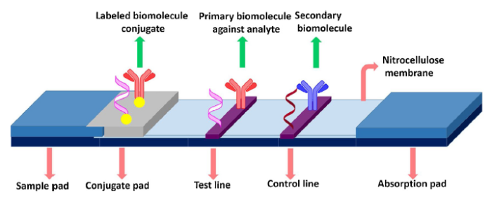
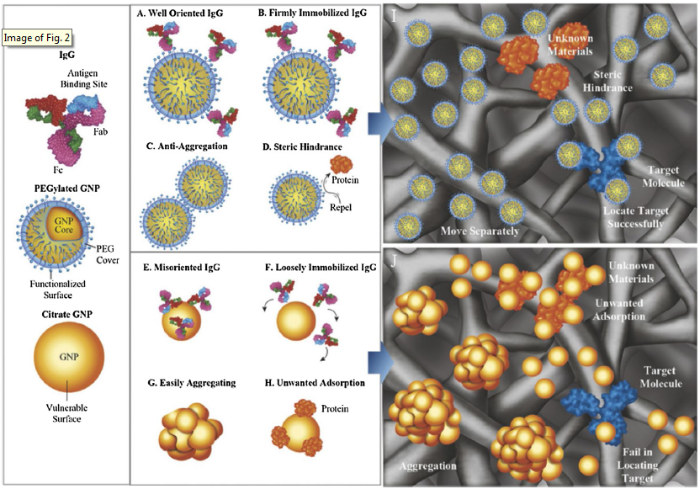
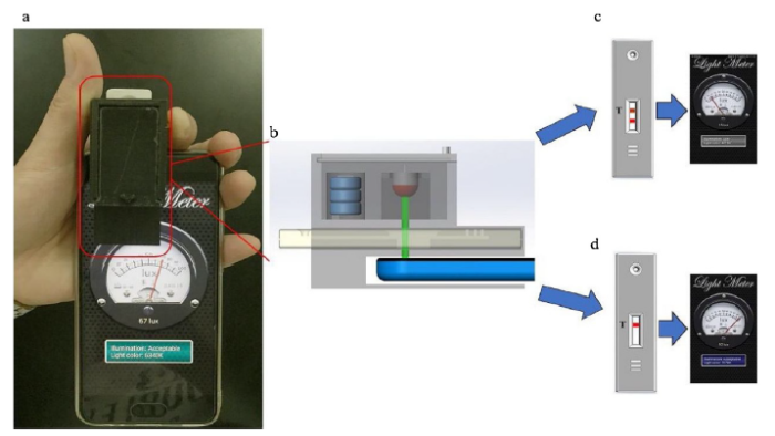
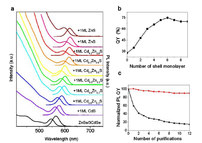
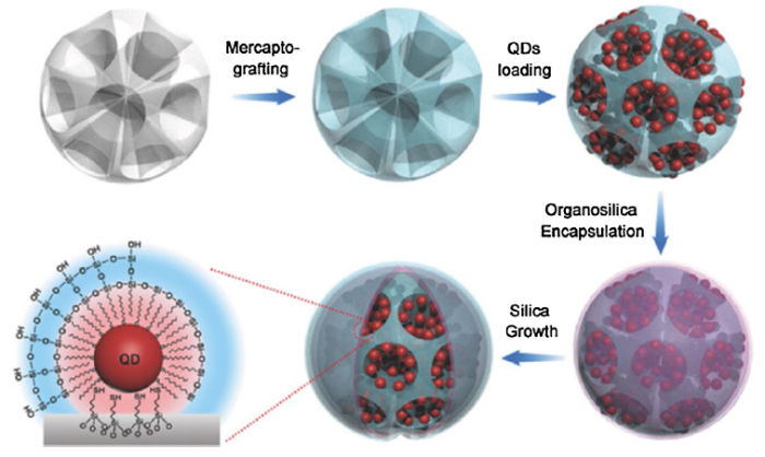
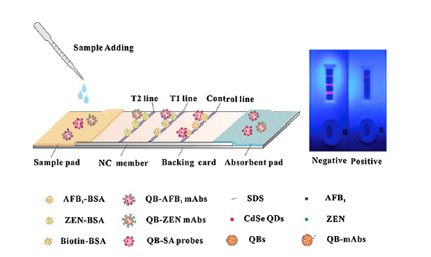
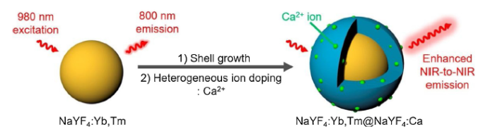
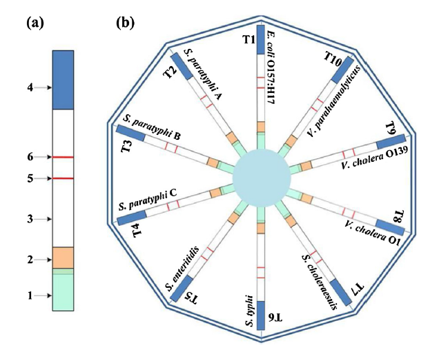


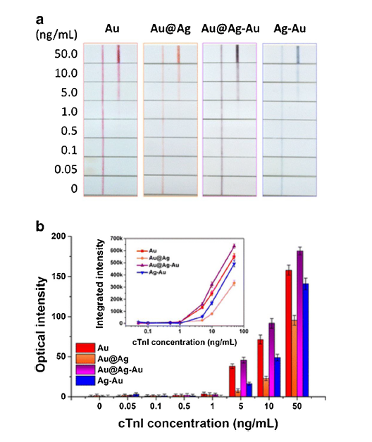

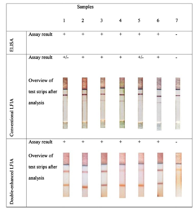
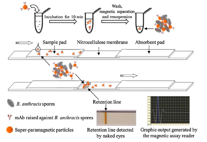
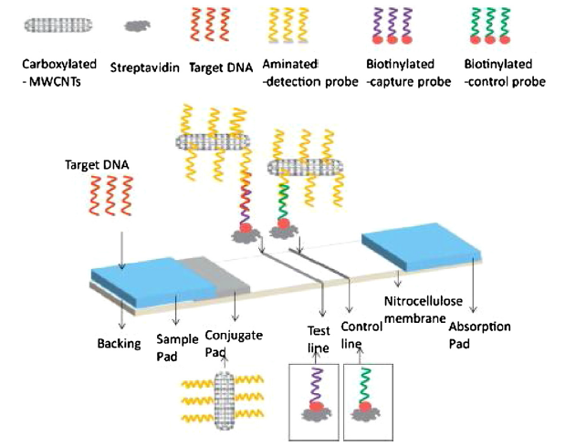


 WeChat