

1. Introduction
As “smart” implants, magnesium (Mg) and its alloys have been turned out to be a promising candidate for orthopedic implant materials or orthopedic appliance [[1], [2], [3]]. The most attractive characteristic of magnesium and its alloy is biodegradability. Providing temporary support during the healing process of bone fracture or defects is the primary purpose of designing degradable biomaterials. But quite apart from it, degradable biomaterials progressively degrade after that [4]. Magnesium is an essential element to human metabolism which naturally exists in bone [5]. Magnesium alloys have favorable biodegradability in the physiological environment and promote new bone formation [6]. A similar Young’s modulus to the human bone can avoid stress shielding. The surrounding natural bones can transfer to the defects and stimulate bone remodeling. However, during screw tightening, the ultimate mechanical strength failed to meet the demand of fixture. It is one of the most challenging issues that the high-purity Mg implants cannot bear the load at loaded bone [7]. Generally, to be a suitable biodegradable orthopedic implant, Mg alloys should be designed with the following properties, including: (1) appropriate mechanical properties for the intended use, (2) a moderate and homogeneous degradation performance and (3) excellent biocompatibility and bioactivity within the human body, which is the critical issues for biomedical applications [[8], [9], [10]].
In order to improve the mechanical properties and corrosion resistance of magnesium alloys, adding alloying elements, microstructure designing and modification are feasible ways. Zn was selected to be the second alloying element, as it can increase strength and ductility. Zr is well known as a useful grain refiner [1]. Rare-earth (RE) elements have a “scavenger effect” on impurities [11]. Neodymium (Nd) and yttrium (Y) are selected to be the alloying elements. Y is of high solid solubility group and Nd is of limited solubility group. Table 1 shows the solubility limits of the main alloying elements in magnesium [9]. Nd is often added to refine the microstructure [[12], [13], [14], [15], [16]]. It is known that Mg alloys with a small amount of Nd exhibited excellent biocompatibility as well [13,17,18]. Y added to magnesium alloys for biodegradable applications due to its beneficial effect on alloy corrosion and strength [19]. Mg alloys can be solidified rapidly and enhanced the corrosion resistance by adding the Y element. The incorporation of oxidized the Y in the surface film was identified as enhancing the corrosion resistance of Mg-Y alloys [20,21].
Table 1 Solubility limits of main alloying elements in magnesium (wt%) [9].
| Element | Solubility |
|---|---|
| Zn | 6.2 |
| Zr | 3.8 |
| Nd | 3.6 |
| Y | 12.4 |
There are several considerations for the choice of elements in the development of magnesium alloys. The human amount, blood serum level, pathophysiology, toxicity levels and a daily allowance of the metal ions are listed in a table by Zheng et al. [1]. The pathophysiology Mg is an activator of many enzymes and a co-regulator of protein synthesis and muscle contraction. Zinc is also an essential component of protein synthesis and energy-producing enzymes, maintains the structural integrity of bio-membrane. As Zn2+ at high concentration was harmful to cell differentiation and mineralization, it should be strictly limited to 4 wt% [9]. Adequate and moderate intake of Mg and Zn is essential for health, the recommended daily intake for adults of Mg is 240-420 mg/day, and zinc is 8-11 mg/day [9,22]. Zr has low systematic toxicity to animals with a daily allowance of 3.5 mg for humans. As alloying, the degradation of products should be non-toxic, tissue absorbable, or excretable via the kidney. Those can be references to be considered during the alloying treatment.
In most of the studies, alloying and surface coating have been used to reduce the degradation rate of Mg. It is believed that the rapid degradation in the physiological environments hampers its clinical use [23]. In the previous study [24], two new Mg-Zn-Zr-Nd and Mg-Zn-Zr-Y alloys were fabricated by casting and hot extrusion. We conducted this study to investigate the practicability of the new Mg-alloys. The physical and mechanical properties were estimated by tensile and fatigue tests. The electrochemical test and immersion test were performed to evaluate the degradation behavior. At last, cytotoxicity, osteogenesis and cell morphology were tested to study the in vitro cytocompatibility.
2. Materials and methods
2.1. Materials and microstructure characterization
The Mg-Zn-Zr-Nd and Mg-Zn-Zr-Y alloys in this study were provided by Institute of Metal Research, Chinese Academy of Sciences. The method can be seen in the previous study [25]. A commercial pure Mg (as-extruded, >99.9 wt%, provided by Institute of Metal Research, Chinese Academy of Sciences, China) was adopted as a control material. The analyzed chemical compositions of the Mg-Zn-Zr-Nd and Mg-Zn-Zr-Y alloys were measured by inductively coupled plasma optical emission spectrometer (ICP-OES) (Optima 8300DV, PerkinElmer, USA). Disc samples of φ10 mm × 2 mm were used for the in vitro study and extract preparation, and φ10 mm × 6 mm were processed for electrochemical tests. All the samples were grounded with SiC paper up to 2000 grit. Then the discs were cleaned in acetone and absolute ethanol by ultrasonic for 10 min each and sterilized with ultraviolet for at least 60 min each side. Before the in vitro cytocompatibility experiments, the discs were sterilized with ethylene oxide.
Microstructure investigation was observed by optical microscopy (OM) (Axio Observer Z1, Zeiss, Germany). The alloy samples were polished and etched in a picric solution (6 g picric, 10 mL acetic, 10 ml water and 70 mL ethanol).
2.2. Mechanical tests
Tensile test specimens of pure Mg, Mg-Zn-Zr-Nd and Mg-Zn-Zr-Y alloys with a diameter of 4 mm and 36 mm gauge length were machined according to ASTM-E8-04 [26] and then successively polished. The tensile tests were performed using the Instron 5982 universal test machine (Instron, USA) at a displacement rate of 1.0 mm/min. At least three specimens were tested in each group. The true stress-strain curve was obtained. The fracture morphologies were taken by SEM (Zeiss Merlin Compact, Zeiss, Germany). Young’s modulus test samples with a diameter of 7-8 mm and 80 mm gauge length was machined according to ASTM E1876-15 and then polished. The fundamental resonant frequency of tested specimens in flexure was recorded by an elastic modulus system (RFDA-HTVP1750-C, IMCE, Belgium) at room temperature.
2.3. Electrochemical test
Electrochemical tests were performed in alpha modified eagle medium (α-MEM, Hyclon, USA) with 10% fetal bovine serum (FBS, Clark, Virginie Ledoyen, USA) in a conventional three-electrode system by using a Reference 600+ Potentiostat/Galvanostat/ZRA automatic laboratory corrosion measurement system (Gamry Instruments, Inc., USA). The water bath method kept the test temperature at 37 °C. A three-electrode cell with a counter-electrode (platinum foil), a reference electrode (a saturated calomel electrode (SCE)) and a sample as a working electrode (0.785 cm2) were used for the test. EIS (electrochemical impedance spectroscopy) and potentiodynamic polarization tests were started after 1800s of initial stabilization. EIS tests were carried out after reaching open circuit potential (OCP) with a 5 mV perturbation. The measuring frequency ranged from 10-2 Hz to 105 Hz. Potentiodynamic polarization tests started from -0.25 V to 0.35 V using a scan rate of 0.5 mV/s. At least three times were repeated in each test to guarantee the reproducibility of the results.
2.4. Immersion tests
The samples for immersion tests were cut into φ10 mm × 2 mm. After ground and polished, corrosion tests consisted of 3 d and 10 d continuous immersion of specimens in α-MEM with 10% fetal bovine serum at 37 °C, 5% CO2. Five samples were prepared in the immersion test. The immersion ratio was 20 mL/cm2. The surface areas of the samples were calculated by measuring through vernier caliper. The inductively coupled plasma mass spectrometry (7800 ICP-MS, Agilent, USA) was used to measure the concentrations of magnesium and alloying element ions (Zn, Zr, Nd, Y). Three immersed samples of each group were cut after mounting them in epoxy resin. The thickness and components of the general corrosion product were measured by SEM and EDS were analyzed. The surfaces of three immersed samples of each group were determined by an X-ray diffractometer (XRD, Shimadzu, XRD-7000). Before and after immersion, five samples were weighed through analytical balance (XS105 DualRange, Mettler Toledo, Switzerland). The balance repeatability (typical) of the analytical balance is 0.01 mg. Each sample was tested three times and the average value was obtained. The corrosion products were removed by chromic acid solution (200 g/L CrO3 + 10 g/L AgNO3) and dried overnight. The corrosion rate was calculated according to ASTM G31-72 [27]. Statistical analysis was performed using one-way ANOVA by turkey’s test, and p < 0.05 was considered statistically significant. After that, pictures were taken by a digital camera.
2.5. Extract preparation
The extracts were used for the in vitro tests. Mg alloy samples were immersed in α-MEM medium with 10% fetal bovine serum for 72 h at 37 °C in a humidified atmosphere of 5% CO2. The immersion ratio was 1.25 mL/cm2 according to ISO 10993 Part 12. The extracts were filtrated and collected. The pH of extracts was determined by a pH meter (PHS-3C, Leici, China), respectively. The extracts were diluted with fresh culture medium for 6 times for cell culture.
2.6. In vitro tests
2.6.1. Cell culture
Murine calvarial preosteoblasts (MC3T3-E1) cells were cultured in α-MEM medium with 10% FBS, 100 U/mL penicillin and 0.1 mg/mL streptomycin and maintained in an incubator at 37 °C, 5% CO2. When the monolayer reached subconfluence, the cells were subcultured by 0.25% trypsinization (Sigma Chemical Co., St. Louis, MO). MC3T3-E1 cells were used to test the cell proliferation, cytotoxicity, adhesion and osteogenesis of pure Mg, Mg-Zn-Zr-Nd and Mg-Zn-Zr-Y alloys.
2.6.2. Cell proliferation and cytotoxicity test
The CCK8 tests were used to evaluate the cytotoxicity of Mg and its alloys by a CCK8 kit (US Everbright Inc., Silicon Valley, USA). Cells were incubated in 96-well cell culture plates (Corning, NY) at 3 × 10³ cells/well. The medium was then replaced with 100 μL of extracts respectively after 24 h to allow attachment, the control groups were replaced by normal culture medium, with five biological replicates per group. The 96-well cell culture plates were incubated in a humidified atmosphere with 5% CO2 at 37 °C for 24 h, 48 h and 72 h. Then 100 μL α-MEM with 10% CCK8 was added to each well, and the 96-well cell culture plates were incubated with CCK8 solution for 4 h at 37 °C. The spectrophotometric absorbance of the samples was measured by a microplate reader (Infinite M200, Tecan, Austria) at 450 nm (490 nm). All tests were repeated three times. Statistical analysis was performed with SPSS 17.0. Differences between groups were analyzed using one-way ANOVA followed by Tukey’s test.
2.6.3. Direct cell viability evaluation
MC3T3-E1 cells were seeded onto the pure Mg, Mg-Zn-Zr-Nd and Mg-Zn-Zr-Y alloys in 24-well plates at a density of 2 × 10⁵/well. The culture media was removed and specimens were fixed with 2.5% glutaraldehyde solution for 4 h at 4 °C after 4 h and 24 h incubation respectively. All the samples were washed 3 times with phosphate buffer solution (PBS, pH = 7.4), and subsequently dehydration in a graded ethanol series (30, 50, 75, 95, 100 vol.%) for 10 min each. The morphologic characteristics of the cells cultured onto the samples’ surface were observed by SEM (Zeiss Merlin Compact, Germany). Three samples were taken for each group.
2.6.4. ALP activity staining
Alkaline phosphatase (ALP) staining was used to test mineralization activity [28], MC3T3-E1 cells were seeded in 6-well plates at 5 × 10⁴ cells in each well. After 24 h of culture, the extract was used to replace the culture medium, and a normal culture medium was used to replace the control group. After 7 and 14 days, cells treated following the manufacturer’s instruction of BCIP/NBT ALP color development kit (Beyotime, China). Purple areas are considered positive, and pictures were taken by a digital camera.
2.7. Fatigue test
Fatigue test samples with a gauge length of 16 mm and a circular cross-section 4 mm in diameter were machined according to ASTM-E466. High cycle fatigue testing was carried out by a computer-controlled servo-hydraulic testing machine (CARE M-3000, Kell Measurement & Control Co., LTD., Tianjin, China). The load control was a push-pull sinusoidal loading with frequency 15 Hz (stress ratio R= -1) in ambient air. The fatigue tests continued until complete failure or until the sample didn’t fail at least 107 cycles.
Mg-Zn-Zr-Nd samples were immersed in Hank’s artificial body fluid [29] for 7, 30, 60, 90 d respectively (the solution was changed every 3 days), then tested with axial sinusoidal loading of 111.5 MPa (fatigue limit in the air condition) and 2 Hz test frequency. The cycles were recorded and the fracture surfaces were examined by SEM.
3. Results
3.1. Chemical composition and microstructural characterization
Analyzed by ICP-OES, the chemical composition of the Mg-Zn-Zr-Nd alloy is: Zn 1.67 wt%, Zr 0.52 wt%, Nd 0.34 wt% and Mg balance. The chemical composition of the Mg-Zn-Zr-Nd alloy is: Zn 1.62 wt%, Zr 0.55 wt%, Y 0.38 wt% and Mg balance.
The optical microstructures of Mg-Zn-Zr-Nd and Mg-Zn-Zr-Y alloy were shown in Fig. 1. The grain size of Mg-Zn-Zr-Nd and Mg-Zn-Zr-Y alloys was measured by the linear intercept method according to ASTM E112-10. There was no significant difference between the average grain sizes of the Mg-Zn-Zr-Nd and Mg-Zn-Zr-Y alloys, which was appropriately 4-5 μm. Grain refinement is often used to increase the corrosion resistance and mechanical properties of Mg alloys. It is hard to distinguish their grain structure by optical microstructures due to the very high density of twins.
Fig. 1.

Fig. 1.
Optical microstructures of Mg-Zn-Zr-Nd alloy (a) and Mg-Zn-Zr-Y alloy (b).
3.2. Mechanical properties
The tensile properties of the RE contained Mg alloys were measured and the true stress-strain curves are illustrated in Fig. 2. The yield strength (YS), ultimate strength (UTS), elongation of Mg-Zn-Zr-Nd and Mg-Zn-Zr-Y alloys were much higher than those of pure Mg. The Young’s modulus of Mg-Zn-Zr-Nd and Mg-Zn-Zr-Y alloy are slightly higher than pure Mg. Results infer that the addition of alloying elements has improved the mechanical properties of the Mg-alloys.
Fig. 2.

Fig. 2.
Typical stress-strain curves and mechanical performances of Mg-Zn-Zr-Nd and Mg-Zn-Zr-Y alloys, taking pure Mg as a control.
The fracture surfaces of the alloys after tensile tests at room temperature were observed by SEM (Fig. 3). Many dimples can be seen on the surfaces. There is no significant difference in the fracture mode of Mg-Zn-Zr-Nd alloy and Mg-Zn-Zr-Y alloy. Both tearing dimples and tearing edges can be discovered in both sections shows the ductile fracture morphology. The UTS and YS of Mg-Zn-Zr-Nd alloy and Mg-Zn-Zr-Y alloy are about 30% and 100% higher than pure Mg, respectively. The elongation of Mg-Zn-Zr-Nd alloy is about 50% higher than that of pure Mg. The UTS of human cortical bone is 70-150 MPa respectively, the elongation is 0-8%, lower than those of the two alloys. Both Young’s modulus of Mg-Zn-Zr-Nd alloy (45.05 ± 0.006 GPa) and Mg-Zn-Zr-Y alloy (44.61 ± 0.017 GPa) are closed to that of human cortical bone (17-20 GPa). In general, Mg-Zn-Zr-Nd alloy exhibited an ideal combination of strength and ductility.
Fig. 3.
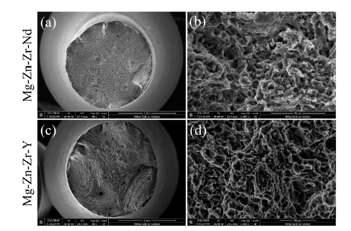
Fig. 3.
Tensile fracture morphologies of Mg-Zn-Zr-Nd alloy (a, b) and Mg-Zn-Zr-Y alloy (c, d).
3.3. Electrochemical corrosion behavior
Fig. 4 shows Nyquist plots and Bode plots of pure Mg, Mg-Zn-Zr-Nd alloy and Mg-Zn-Zr-Y alloy immersed in α-MEM with 10% FBS solutions at 37 °C. Two capacitive loops at high and medium frequencies were exhibited in pure Mg, and an inductive loop was shown at low frequency (Fig. 4(a)). There was a capacitive loop and an inductive loop at high and low frequency respectively in the Nyquist plot of Mg-Zn-Zr-Nd alloy and Mg-Zn-Zr-Y alloy. The capacitive loop showed the spectra and the diameter of Mg-Zn-Zr-Nd and Mg-Zn-Zr-Y alloy were similar (Fig. 4(a)) which indicated that the corrosion performance of the samples is similar. As is known to all, in the Nyquist plot, the larger the diameter of the capacitive loop, the stronger the corrosion resistance in the solution [30]. Pure Mg showed the largest diameter of the Nyquist loop, indicated that pure Mg had the lowest corrosion rate. The inductive loop at low frequency implied the breakdown of corrosion film caused by localized corrosion [31]. The resistance of Mg-Zn-Zr-Nd and Mg-Zn-Zr-Y alloy in the Bode diagram at low frequency is almost the same (Fig. 4(b)).
Fig. 4.

Fig. 4.
Electrochemical corrosion behaviors of the experimental materials in α-MEM solution with 10% FBS at 37 °C: (a) Nyquist plots; (b) Bode plots; (c) potentiodynamic polarization curves; (d) electrochemical data. Ecorr represents corrosion potential and icorr represents corrosion current density (n = 3, * p < 0.05).
Fig. 4(c) shows the electrochemical polarization curves of Mg-Zn-Zr-Nd and Mg-Zn-Zr-Y alloys in the α-MEM solution with 10% FBS solutions. The addition of the alloy elements Zn, Zr, Nd, Y improved the corrosion potential of the alloys and the polarization curves of Mg-Zn-Zr-Nd and Mg-Zn-Zr-Y were similar. Corrosion potential (Ecorr) and corrosion current density (icorr) were calculated directly from the polarization plots through the Tafel region extrapolation (Fig. 4(d)). There were no significant differences between the corrosion current density of Mg-Zn-Zr-Nd, Mg-Zn-Zr-Y alloys and pure Mg (p > 0.05).
3.4. Immersion corrosion behavior
Immersion tests were performed to observe a relative long-term corrosion behavior. Fig. 5 shows the surface features of the corroded pure Mg, Mg-Zn-Zr-Nd and Mg-Zn-Zr-Y alloys after immersion tests in α-MEM with 10% FBS solutions at 37 °C 5% CO2 for 3 and 10 day, respectively. From the appearance of the sample surface, Mg-Zn-Zr-Y alloy was more severely corroded than pure Mg and Mg-Zn-Zr-Nd alloy. The corrosion rate was calculated according to the following equation (ASTM G31-72):
Fig. 5.

Fig. 5.
(a) Corroded surface photographs (corrosion products were removed); (b) corrosion rate of pure Mg, Mg-Zn-Zr-Nd and Mg-Zn-Zr-Y alloy after immersion in α-MEM with 10% FBS solution at 37 °C 5% CO2 for 3 d and 10 d, respectively (n = 5, *p < 0.05); (c) ion concentration in immersion test of pure Mg, Mg-Zn-Zr-Nd and Mg-Zn-Zr-Y alloy according to ICP-MS test.
where C is the corrosion rate (mmpy), coefficient K is 8.76 × 10⁴, W is the weight loss (g), A is the exposed sample area (cm2), T is the exposure time (h), and D is the density of the material (g/cm3) (ρMg: 1.733 g/cm3, ρMg-Zn-Zr-Nd: 1.776 g/cm3, ρMg-Zn-Zr-Y: 1.776 g/cm3). The corrosion rates of the alloys at different times are shown in Fig. 5(b). On day 3, the corrosion rate shows in an order of Mg-Zn-Zr-Y alloy > Mg-Zn-Zr-Nd alloy > pure Mg, but pure Mg shows no significant difference with Mg-Zn-Zr-Nd alloy. On day 10, all the corrosion rates are decreased, but the tendency of average values hasn’t changed. There is a significant difference between Mg-Zn-Zr-Y alloy and Mg-Zn-Zr-Nd alloy. The Mg2+ concentration and the corrosion rate that calculated according to the immersion test are consistent. Localized corrosion could be seen on all the samples (Fig. 5 (a)).
Fig. 6 shows the EDS spectra of the cross-section of three materials after immersion in α-MEM with 10% FBS solutions for 72 h with corresponding SEM images. Mg is the major element of the non-degraded substrate and the corrosion pits of pure Mg are wide and shallow. Comparatively, the pits of Mg-Zn-Zr-Nd are small and slightly more in depth, while those of Mg-Zn-Zr-Y are wide and deep. All the samples were rich in Ca, P and O. Moreover, Zr was detected in the corrosion layers of the alloys. Calcium and phosphorus are necessary to maintain bone mass and essential to cell growth. The thicknesses of corrosion products on pure Mg were 35-40 μm after immersion, whereas those on Mg-Zn-Zr-Nd and Mg-Zn-Zr-Y alloys were 10-14 μm and 8-10 μm, respectively.
Fig. 6.

Fig. 6.
EDS mapping of cross-sections of corrosion products after immersion tests in solution for 3 days: (a) pure Mg; (b) Mg-Zn-Zr-Nd alloy; (c) Mg-Zn-Zr-Y alloy.
Fig. 7 presents the XRD patterns of Mg-Zn-Zr-Nd and Mg-Zn-Zr-Y alloys after 3 d and 10 d immersion in α-MEM with 10% FBS. All the samples exhibited α-Mg as a single phase. After 3 d of immersion, there was no significant difference among all samples. Ca3(PO4)2 and Mg3(PO4)2 were detected on surfaces of 10 d samples. It appeared that only calcium phosphate and magnesium phosphate were deposited on the surfaces. The presence in the corrosion products or conversion films formed on Mg alloys improved their corrosion resistance in an aggressive environment.
Fig. 7.
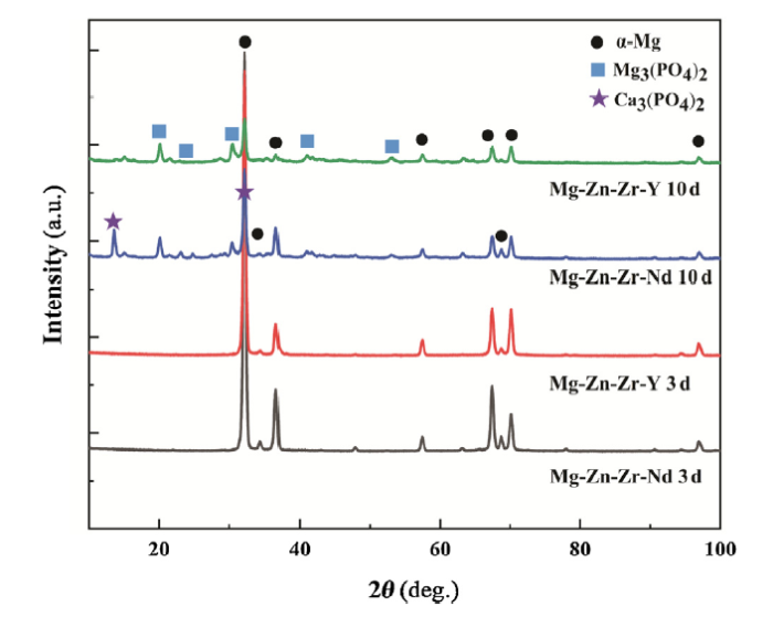
Fig. 7.
XRD patterns of Mg-Zn-Zr-Nd and Mg-Zn-Zr-Y alloys after 3 d and 10 d immersion in α-MEM with 10% FBS.
3.5. pH of extracts
Fig. 8 shows the pH value and ion concentration released from pure Mg, Mg-Zn-Zr-Nd alloy and Mg-Zn-Zr-Y alloy in α-MEM solution with 10% FBS at 37 °C in a 5% CO2 humidified air. The pH values of Mg-Zn-Zr-Nd and Mg-Zn-Zr-Y alloy extracts were similar, while both were higher than pure Mg (p < 0.05).
Fig. 8.

Fig. 8.
pH values of pure Mg, Mg-Zn-Zr-Nd and Mg-Zn-Zr-Y alloys in α-MEM solution with 10% FBS at 37 °C 5% CO2.
3.6. In vitro tests
3.6.1. Cell proliferation and cytotoxicity
The optical densities of MC3T3-E1 in different extracts were measured by the CCK8 test (Fig. 9(a)). On 24 h, there was no significant difference among all the groups (p > 0.05). On 48h, the absorbance of Mg-Zn-Zr-Y group showed significantly lower than Mg-Zn-Zr-Nd (p < 0.001) and pure Mg group (p < 0.01). On 72 h, all the extracts were significantly lower than control group. In Fig. 9(b) the relative growth rate (RGR) of MC3T3-E1 cells in extracts of Mg-Zn-Zr-Nd alloy and Mg-Zn-Zr-Y alloy and pure Mg were assessed. RGR is calculated as follows:
Fig. 9.

Fig. 9.
(a) Optical density of MC3T3-E1 cells in extraction medium of experimental materials measured by CCK8 test and (b) relative growth rate (RGR) and cytotoxicity level at different detection period (n = 5, *p < 0.01, **p < 0.001).
ODe is the average OD value of the experimental groups; ODc is the average OD value of the control group. The cell toxicity grade (CTG) is obtained according to the standard United States Pharmacopeia [32].
The results showed that CTG of the Mg-Zn-Zr-Nd alloy was in grade 0 which represents no toxicity. From 24 h to 72 h of incubation, Mg-Zn-Zr-Y alloy showed grade 0 to 1(no toxicity).
3.6.2. Direct cell viability evaluation
The morphologies of MC3T3-E1 cultured on Mg-Zn-Zr-Nd, Mg-Zn-Zr-Y alloys and Pure Mg for 4 h and 24 h are shown in Fig. 10. After 4 h culture, cells attached to all samples. The cells on Mg-Zn-Zr-Nd alloy are of lager quantity, osteoblastic shape and spread well, while the cells on pure Mg and Mg-Zn-Zr-Y alloy are mostly round-shaped. After 24 h culture, cells attached and spread well on all samples. The polygonal osteoblastic shape with finger-like protrusions can be seen. However, it was observed that the cells spread more extensions of filopodia and abundant cytoplasm on the Mg-Zn-Zr-Nd alloy surface than on pure Mg and Mg-Zn-Zr-Y alloy at high magnification.
Fig. 10.

Fig. 10.
Morphologies of MC3T3-E1 cells cultured on pure Mg, Mg-Zn-Zr-Nd and Mg-Zn-Zr-Y alloys for 4 h and 24 h: (a) pure Mg for 4 h; (b)Mg-Zn-Zr-Nd for 4 h; (c) Mg-Zn-Zr-Y for 4 h; (d) pure Mg for 24 h; (e) Mg-Zn-Zr-Nd for 24 h; (f) Mg-Zn-Zr-Y for 24 h.
3.6.3. ALP activity
Alkaline phosphatase (ALP) plays an essential role in mineral deposition and generally considered as an osteogenic marker [33]. Fig.11 shows the ALP staining of MC3T3 cells cultured with pure Mg, Mg-Zn-Zr-Nd alloy and Mg-Zn-Zr-Y alloy extracts for 7 days and 14 days, respectively. After 7 d and 14 d of cell culture, the cells were stained purple. On day 7, the staining of the Mg-Zn-Zr-Nd alloy group was darker than pure Mg group, while the color of the Mg-Zn-Zr-Y group could barely be seen. While on day 14, the staining of pure Mg group was like the Mg-Zn-Zr-Nd alloy group, but only tiny spots of purple staining could be seen in the Mg-Zn-Zr-Y alloy group. Alkaline phosphatase staining was used to observe the effect of the extract on the activity of alkaline phosphatase.
Fig. 11.
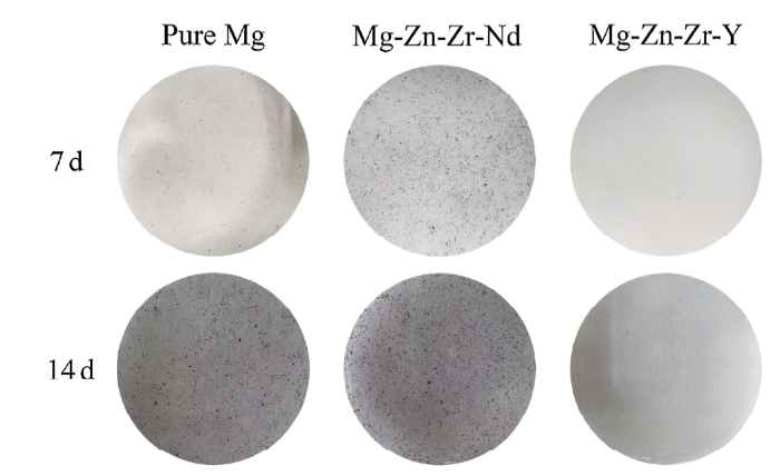
Fig. 11.
ALP staining of MC3T3-E1 cells cultured with pure Mg, Mg-Zn-Zr-Nd alloy and Mg-Zn-Zr-Y alloy extracts for 7 d and 14 d, respectively.
3.7. Fatigue limits
Fig. 12(a) shows the S-N curve of Mg-Zn-Zr-Nd alloy tested in air. The fatigue limit of Mg-Zn-Zr-Nd alloy at 107 cycles was 107.5 MPa. Fig. 12(b) shows that the cycles of failure decreased as the immersion time increased. The crack nucleation region of the unsoaked Mg-Zn-Zr-Nd alloy was relatively flat (Fig. 13(a-d)). Fig. 13(e, i, m, q) shows the overall fatigue fracture surfaces after Mg-Zn-Zr-Nd alloy samples immersed for 7, 30, 60, 90 days, respectively. The failure of the Mg-Zn-Zr-Nd alloy could be ascribed to the formation of the corrosion pits, resulting in the crack initiation. As the more time immersed in simulated body fluid, the more corrosion initiation sites in the rim region could be found on the samples.
Fig. 12.
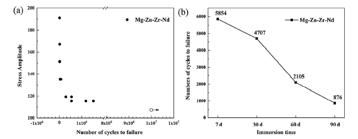
Fig. 12.
(a) S-N curves of Mg-Zn-Zr-Nd alloy in air (data points with arrows indicate that the specimens did not fail) and (b) cycles of failure after immersed in Hank’s solution for 7 d, 30 d, 60 d, 90 d, respectively.
Fig. 13.
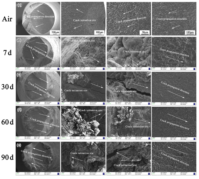
Fig. 13.
SEM images showing the fatigue fracture morphologies of Mg-Zn-Zr-Nd alloy in air (failure at 1184616 cycles, under the stress amplitude of 115 MPa and test frequency of 15 Hz) (a), immersed in Hank’s solution for 7 d (failure at 5854 cycles, under the stress amplitude of 111.4 MPa and test frequency of 2 Hz) (b), immersed in Hank’s solution for 30 d (failure at 4707 cycles, under the stress amplitude of 111.4 MPa and test frequency of 2 Hz) (c), immersed in Hank’s solution for 60 d (failure at 2105 cycles, under the stress amplitude of 111.4 MPa and test frequency of 2 Hz) (d), immersed in Hank’s solution for 90 d (failure 876 cycles, under the stress amplitude of 111.4 MPa and test frequency of 2 Hz) (e).
The crack initiation zone and the crack propagation direction were marked in Fig. 13. Fig. 13(a-d) shows the typical high-cycle fatigue fracture surfaces of the Mg-Zn-Zr-Nd alloy tested in the air. With the increasing dipping time in Hank’s solution, the corrosion was aggravated, while the failure cycles decreased.
4. Discussion
4.1. Mechanical properties of Mg-Zn-Zr-Nd and Mg-Zn-Zr-Y alloys
The Mg-Zn-Zr-Nd and Mg-Zn-Zr-Y alloys in present study were extruded at 390 °C at an extrusion ratio of 64. It causes element diffusion and microstructure changes [34,35]. The enhancement of strength is mainly caused by the introduction of residual stress and the contradictory effect of microscopic refinement [36]. The fracture mode of Mg-Zn-Zr-Nd and Mg-Zn-Zr-Y alloys was mainly trans-granular ductile fracture, there are many dimples and tear ridges on the fracture surface (Fig. 3). The results are consistent with the elongation of both alloys. No apparent second phase was detected in both alloys. The improved stress of alloys originated from the homogeneous microstructure and the addition of Zn, Zr and RE elements. The addition of Zn, Zr, Nd and Y are helpful to the grain refinement by inhibiting the impurity and oxygen which would enhance the tensile strength, hardness and ductility. When zinc content increased, the decrease in the basal texture intensity may affect the yield strength reduction and the elongation increase. However, it should be limited under the maximum solubility(6.2 wt%), or it would form eutectic and pores phases to reduce the strength [37]. As everyone knows, Zr is a grain refiner. Saha et al. [38] the addition of amounts of Zr (0.25 and 1.0 wt%) resulted in grain refinement and an increase in mechanical properties. In our study, the Zr element was added at approximately 0.5 wt% which could be considered as an appropriate amount. Nd improves the mechanical properties of magnesium alloy mainly through the following three mechanisms. Firstly, Nd has a good grain refinement effect. Secondly, is the strengthening effect of the second phase. Finally, the solid-soluble Nd atoms can activate the non-base slip and reduce the stacking fault energy of the alloy. Compared with Nd, the addition of Y enhances strength, while decreases elongation [39].
The ratio of YS and UTS is named yield ratio (σys/σb). The lower the yield ratio is, the less likely plastic instability occur. The yield ratio of Mg-Zn-Zr-Nd and Mg-Zn-Zr-Y alloys are 0.856 and 0.885, respectively, which represent that the Mg-Zn-Zr-Nd alloy is more stable and more suitable for medical materials.
4.2. Corrosion behaviors of Mg-Zn-Zr-Nd and Mg-Zn-Zr-Y alloys
Mg-Zn-Zr-Nd alloy, Mg-Zn-Zr-Y alloys and pure Mg exhibited similar cathodic current densities, suggesting similar corrosion resistance. In Nyquist plots, as we all know, the larger loop means better corrosion resistance [40]. Hence pure Mg was more anti-corrosive than Mg-Zn-Zr-Nd and Mg-Zn-Zr-Y alloys, corresponding to the polarization curves, as illustrated in Fig. 4. As the corrosion potential and Nyquist loop of Mg-Zn-Zr-Nd and Mg-Zn-Zr-Y alloys were nearly the same, we could infer that Nd and Y were not the main factors that caused the difference of the alloys.
The corrosion potentials of the samples show an order as follows: Mg-Zn-Zr-Nd ≈ Mg-Zn-Zr-Y < pure Mg, which means that pure Mg may be most probably to be corroded in the α-MEM solution. The corrosion current density (icorr) was obtained by the polarization curve, and Fig. 4 shows that icorr of the three samples did not demonstrate significant differences. Present studies discovered that the corrosion products of Mg alloy are composed of three layers: an loose outer layer (Mg(OH)2), a dense thin intermediate MgO region and hydrated inner MgO-Mg(OH)2 layer [41]. The chloride ion of the α-MEM solution is much higher than 30 mM. Consequently, Mg(OH)2 reacted continuously in the α-MEM solution and accelerated the corrosion behavior. Since no protective layer was formed on the surface at the beginning, all the samples corroded faster at the initial stage. While pH < 11.5, the reactions of magnesium alloys and the aqueous solution facilitate [42]. So, part of the corrosion layer is destroyed, the defect is further generated and the corrosion is intensified.
The degradation behavior of Mg in the liquid environment can be summarized as the following equation [43]:
The cathodic hydrogen evolution was reflected by the cathodic polarization curve through water reduction on the platinum electrode. The anodic polarization curve represented the dissolution of magnesium on saturated calomel electrode [44]. As the anodic and cathodic branches were not symmetrical, corrosion current density (icorr) and the corrosion potential (Ecorr) were calculated by the cathodic branch of Tafel extrapolation [13,[45], [46], [47], [48], [49]]. From the thermodynamics point, the addition of Zn, Zr and Nd/Y into Mg stabilized the matrix.
The corrosion resistance was not enhanced mainly because of galvanic corrosion. One of the factors that may affect corrosion is the standard electrode potential. Even Nd (-2.43 V) and Y (-2.37 V) have a close electrochemical potential to Mg (-2.37 V), Zn (-0.76 V) and Zr (-1.53 V) can accelerate the corrosion behavior by promoting the galvanic corrosion. Besides, a large amount of Ca, P, O were detected in the corrosion product layer among all three samples by EDS. Mg reacted with water and formed unstable Mg(OH)2. Mg(OH)2 soon reacted with Cl- in the culture medium and more detectable Mg2+ released. While, as the OH- producing continuously, Mg2+ and Ca2+ reacted with PO43- and formed insoluble Mg3(PO4)2 and Ca3(PO4)2 which are also detected in XRD tests. Mg3(PO4)2 and Ca3(PO4)2 formed a dense protective layer on the samples’ surfaces.
Calcium and phosphorus formed a protective layer and are essential for cell attachment which helps the protein absorption. The existence of calcium and phosphorus may promote the adsorption of proteins on biomaterial surfaces [50].
4.3. Cytobiocompatibility of Mg-Zn-Zr-Nd and Mg-Zn-Zr-Y alloys
The cytocompatibility of Mg-Zn-Zr-Nd and Mg-Zn-Zr-Y alloys was investigated with cell proliferation, cytotoxicity test, direct cell viability evaluation and ALP activity test. Once Mg or Mg alloy contact with the aqua environment, the hydrogen gas evolves from the sample surface, which may decline the cell adhesion and the cell proliferation process [36].
As the direct cell adhesion experiments could rapidly react to the viability and cytotoxicity of the cells, we observed the morphology of the cells by SEM on 4 h and 24 h in our study. The alternation of pH value can regulate the survival, growth and metabolism of cells. Khajah et al. [52] studied the alkaline pH of extracellular from 7.7 to 8.3, and the cell morphology get shrinkage and spherical appearance. In our study, MC3T3-E1 cells on pure Mg and Mg-Zn-Zr-Y alloy showed spherical appearance at 4 h, however, they spread better at 24 h. Grillo et al. [51]discovered that cells had a better tolerant of gradual alterations in pH and corrosion products in direct assays than in indirect assays. They also discovered that the combination of different concentrations of Mg2+ and Zn2+ could show synergistic or joint antagonist effects. The high viability of Mg-Zn-Zr-Nd alloy may vary because of the advisable combination Mg2+ and Zn2+. Zn ions are involved in many metalloenzymes and proteins, including alkaline phosphatase (ALP) [52,53] and can promote osteoblast cell proliferation and differentiation and thus bone formation [[54], [55], [56]]. However, studies have shown that a high concentration of zinc can induce apoptosis of different cell types and tissues. The concentration of Zn2+ of Mg-Zn-Zr-Y alloy in the immersion test was about 2-folded of Mg-Zn-Zr-Nd alloy, the lower viability of Mg-Zn-Zr-Y alloy might be ascribed to the relatively high Zn2+ concentration.
The mechanical properties and corrosion performance are improved by the addition of Nd [[13], [14], [15], [16]]. Furthermore, the addition of a small amount of Nd also exhibited excellent biocompatibility, which had already been reported [13,17,18]. Also, Nd3+ promoted the differentiation and formation of mineralized matrix nodules of osteoclasts at concentrations of 1 × 10⁻⁸ mol/L and 1 × 10⁻⁵ mol/L, respectively [57]. It is consistent with the excellent performance of cell proliferation and osteogenesis in the Mg-Zn-Zr-Nd group. On the contrary, the biosafety of the Y element is still controversial. In the present study, the cytotoxicity levels were Grade 0, which means no toxicity. On the time point of 7 days, the ALP activity shows a relationship as follows: Mg-Zn-Zr-Nd > pure Mg > Mg-Zn-Zr-Y. While on day 14, the ALP activity of pure Mg and Mg-Zn-Zr-Nd alloy are almost the same, but remarkably few tiny spots can be seen for the Mg-Zn-Zr-Y group. Zhang et al. [58] found that the concentration and culture time are vital factors for switching the biological effects of Y3+ from toxicity to activity, or from down-regulation to up-regulation. Hirano et al. [59] suggested that the accumulate of Y3+ would lead to liver toxicity. So, cells are sensitive to the Y element, which may influence the proliferation and mineralization.
Lee et al. [60] indicated that Zr could promote the proliferation of osteoblasts at a low concentration, and inhibit the proliferation at a high concentration. In contrast, Yamamoto et al. [61] indicated that when Zr ions were lower than 10-3 M/L, it did not cause toxicity for L-929 and MC3T3-E1. Therefore, Zr is not an essential factor affecting cell proliferation and differentiation.
4.4. Fatigue strength
As the common mechanical properties and cytocompatibility of Mg-Zn-Zr-Nd alloy are superior to pure Mg and Mg-Zn-Zr-Y, the fatigue tests were conducted before and after immersed in Hank’s solution to ensure the safety of long service. Studies [62,63] showed the fatigue limits of Mg-1Ca, Mg-2Zn-0.2Ca were around 70 MPa, HP-Mg was 52 MPa, as cast AZ91D was 50 MPa, as extruded WE43 was 110 MPa. The fatigue strength of the Mg-Zn-Zr-Nd alloy was 119 MPa for 5.42 × 10⁵ cycles, which were higher than the alloys above and bone fatigue strength (23-30 MPa) [63]. However, the biomaterials are designed to use in tissues, and the human body is a complicated aqueous environment. What’s more, the complex cyclic loading on the implants in the human body can further accelerate the corrosion. So further studies are still needed to investigate the corrosion and fatigue properties.
5. Conclusions
In the present study, two newly developed biodegradable Mg alloys Mg-1.7Zn-0.5Zr-0.3Nd (Mg-Zn-Zr-Nd) and Mg-1.6Zn-0.6Zr-0.4Y (Mg-Zn-Zr-Y) were investigated. The results can be concluded as follows:
(1) The alloying improved the strength and ductility of Mg-Zn-Zr-Nd and Mg-Zn-Zr-Y alloy. The enhanced Young’s modulus is close to that of human bone. Mg-Zn-Zr-Nd alloy showed the best combination of strength and ductility.
(2) The corrosion resistance of Mg-Zn-Zr-Nd and Mg-Zn-Zr-Y alloys were not enhanced obviously. The initial corrosion resistance of Mg-Zn-Zr-Nd and Mg-Zn-Zr-Y alloys was lower than pure Mg, which might because of the galvanic corrosion. As the corrosion time increases, Mg-Zn-Zr-Nd alloy showed a similar the corrosion rate of pure Mg, which mainly because the Ca3(PO4)2 and Mg3(PO4)2 formed on the surfaces of the samples increase the corrosion resistance. The metal ion concentrations were consistent with corrosion rate.
(3) The Mg-Zn-Zr-Nd and Mg-Zn-Zr-Y alloy extracts showed no toxicity and Mg-Zn-Zr-Nd extract enhanced the osteogenesis. MC3T3-E1 cells adhered at early 4 h and presented polygonal shape, which showed better biocompatibility than pure Mg and Mg-Zn-Zr-Y alloy
In summary, even though the corrosion resistance of Mg-Zn-Zr-Nd and Mg-Zn-Zr-Y alloys were not improved obviously, both mechanical properties have been enhanced significantly. Comparatively, Mg-Zn-Zr-Nd alloy exhibits high potential to be used as orthopedic implant materials under the condition that the corrosion rate should be carefully controlled.
Acknowledgments
This work was financially supported by the National Natural Science Foundation of China (No. U1737102), the Young Elite Scientists Sponsorship Program by China Association for Science and Technology (No. 2017QNRC001), the Shenyang Key R&D and Technology Transfer Program (No. Z18-0-027) and the Key Program of China on Biomedical Materials Research and Tissue and Organ Replacement (Nos. 2016YFC1101804 and 2016YFC1100604).
Reference
DOI
URL
PMID
[Cited within: 1]

As bioabsorbable materials, magnesium alloys are expected to be totally degraded in the body and their biocorrosion products not deleterious to the surrounding tissues. It's critical that the alloying elements are carefully selected in consideration of their cytotoxicity and hemocompatibility. In the present study, nine alloying elements Al, Ag, In, Mn, Si, Sn, Y, Zn and Zr were added into magnesium individually to fabricate binary Mg-1X (wt.%) alloys. Pure magnesium was used as control. Their mechanical properties, corrosion properties and in vitro biocompatibilities (cytotoxicity and hemocompatibility) were evaluated by SEM, XRD, tensile test, immersion test, electrochemical corrosion test, cell culture and platelet adhesion test. The results showed that the addition of alloying elements could influence the strength and corrosion resistance of Mg. The cytotoxicity tests indicated that Mg-1Al, Mg-1Sn and Mg-1Zn alloy extracts showed no significant reduced cell viability to fibroblasts (L-929 and NIH3T3) and osteoblasts (MC3T3-E1); Mg-1Al and Mg-1Zn alloy extracts indicated no negative effect on viabilities of blood vessel related cells, ECV304 and VSMC. It was found that hemolysis and the amount of adhered platelets decreased after alloying for all Mg-1X alloys as compared to the pure magnesium control. The relationship between the corrosion products and the in vitro biocompatibility had been discussed and the suitable alloying elements for the biomedical applications associated with bone and blood vessel had been proposed.
DOI
URL
PMID
[Cited within: 1]
Zinc is required for the activity of > 300 enzymes, covering all six classes of enzymes. Zinc binding sites in proteins are often distorted tetrahedral or trigonal bipyramidal geometry, made up of the sulfur of cysteine, the nitrogen of histidine or the oxygen of aspartate and glutamate, or a combination. Zinc in proteins can either participate directly in chemical catalysis or be important for maintaining protein structure and stability. In all catalytic sites, the zinc ion functions as a Lewis acid. Researchers in our laboratory are dissecting the determinants of molecular recognition and catalysis in the zinc-binding site of carbonic anhydrase. These studies demonstrate that the chemical nature of the direct ligands and the structure of the surrounding hydrogen bond network are crucial for both the activity of carbonic anhydrase and the metal ion affinity of the zinc-binding site. An understanding of naturally occurring zinc-binding sites will aid in creating de novo zinc-binding proteins and in designing new metal sites in existing proteins for novel purposes such as to serve as metal ion biosensors.
DOI
URL
PMID
[Cited within: 1]
Zinc is an important mineral that is required for normal bone development. However, the direct effects of zinc on the mineralization of bone cells of human origin are not clear. The objective of this study was to determine the effects of zinc on the differentiation of SaOS-2 human osteoblastlike cells and the formation of mineralized bone nodules. Cells were cultured for 8 d and then transferred to zinc-free medium and treated with varying concentrations (0-50 microM) of zinc. Alkaline phosphatase (ALP) activity was used as a measure of osteoblast differentiation, and bone nodules were detected by von Kossa staining. After 4, 6, and 8 d of treatment, zinc increased ALP activity at 1 and 10 microM, but decreased activity at 50 microM. After 9 d of treatment, zinc increased both the number and area of mineralized bone nodules at low concentrations (1 and 10 microM), but decreased both at higher concentrations (25 and 50 microM). These findings demonstrate that zinc has biphasic effects on the differentiation and mineralization of human osteoblast-like cells.
DOI
URL
PMID
[Cited within: 1]
This study investigated the resorptive activity of osteoclasts on tricalcium phosphate (TCP), zinc-containing tricalcium phosphate (ZnTCP) and magnesium-containing tricalcium phosphate (MgTCP) ceramics in different Zn- or Mg-containing culture media. On the TCP ceramic, an increase in Zn ions in the culture medium within the range between 0.3 and 6.8 ppm significantly induced an increase in osteoclast apoptosis and a decrease in actin ring formation. However, even a high level of Mg ions up to 100 ppm in the culture medium was unlikely to induce an increase in osteoclast apoptosis. Mg ions in the MgTCP ceramics have no effect on osteoclast apoptosis and actin ring formation. There was almost no significant difference in osteoclast apoptosis and actin ring formation between ZnTCP and MgTCP ceramics which have the same solubility and dissolution rates. It is indicated that only an increase in Zn level outside resorption lacuna has an inhibitory effect on osteoclast resorption and that an increase in Zn level inside resorption lacuna could not influence the osteoclast activity.
DOI
URL
PMID
[Cited within: 1]
Recent years have witnessed a rapid increase in the use of zirconium (Zr)-containing compounds in artificial internal organs. Examples include dental implants and other restorative practices, total knee and hip replacement, and middle-ear ossicular chain reconstruction. In nephrological practice, Zr-containing sorbents have been used in hemofiltration, hemodialysis, peritoneal dialysis, and in the design and construction of wearable artificial kidneys. Zr compounds continue to be widely and extensively used in deodorant and antiperspirant preparations. In the public health arena, Zr compounds have been studied or used in controlling phosphorus pollution and in the reclamation of poison and bacteria-contaminated water. Experimental and clinical studies support the general consensus that Zr compounds are biocompatible and exhibit low toxicity. Reports on possible Zr-associated adverse reactions are rare and, in general, have not rigorously established a cause-and-effect relationship. Although publications on the use of Zr compounds have continued to increase in recent years, reports on Zr toxicity have virtually disappeared from the medical literature. Nevertheless, familiarity with, and continued vigilant monitoring of, the use of these compounds are warranted. This article provides an updated review on the biomedical use of Zr compounds.
DOI
URL
PMID
[Cited within: 1]
UNLABELLED: The dynamic loading in human body, along with the corrosive body fluid, presents a great challenge for the practical use of biodegradable magnesium implants. In this study, a high purity magnesium (99.99wt.%) and two typical promising biodegradable magnesium alloys (binary Mg-1Ca and ternary Mg-2Zn-0.2Ca) were chosen as the experimental materials. Their dynamic mechanical performances were comparatively evaluated by carrying out fatigue tests in air and in simulated body fluid (SBF). The fatigue strengths of HP-Mg, Mg-1Ca and Mg-2Zn-0.2Ca were all around 90MPa in air, however, they decreased to 52MPa, 70MPa and 68MPa in SBF at 4x10(6)cycles, respectively. The fatigue cracks initiated from the microstructural defects when tested in air, but nucleated from surface corrosion pits when tested in SBF. Cyclic loading significantly increased the corrosion rates of all the experimental materials compared to that in static SBF. Moreover, based on our findings, the fatigue failure processes and interactions between material, corrosion and cyclic loading were systematically discussed. STATEMENT OF SIGNIFICANCE: Fatigue strength and life are vital parameters to the design of metallic implant devices. For the corrosion fatigue of biomedical magnesium alloys, we reported the corrosion fatigue behavior of AZ91D and WE43 in SBF (Acta Biomaterialia, 6 (2010) 4605-4613), and till now there is no other reports to our knowledge. We spent 3years to finish the fatigue testing and get S-N curves for three more magnesium biomaterials, and our significant finding is that the fatigue strengths of HP-Mg, Mg-1Ca and Mg-2Zn-0.2Ca are all around 90MPa in air but 52MPa, 70MPa and 68MPa in SBF at 4x10(6)cycles, which will provide the first-hand data for the future magnesium implants design.
DOI
URL
PMID
[Cited within: 2]
Magnesium alloys have been recently developed as biodegradable implant materials, yet there has been no study concerning their corrosion fatigue properties under cyclic loading. In this study the die-cast AZ91D (A for aluminum 9%, Z for zinc 1% and D for a fourth phase) and extruded WE43 (W for yttrium 4%, E for rare earth mischmetal 3%) alloys were chosen to evaluate their fatigue and corrosion fatigue behaviors in simulated body fluid (SBF). The die-cast AZ91D alloy indicated a fatigue limit of 50MPa at 10(7) cycles in air compared to 20MPa at 10(6) cycles tested in SBF at 37 degrees C. A fatigue limit of 110MPa at 10(7) cycles in air was observed for extruded WE43 alloy compared to 40MPa at 10(7) cycles tested in SBF at 37 degrees C. The fatigue cracks initiated from the micropores when tested in air and from corrosion pits when tested in SBF, respectively. The overload zone of the extruded WE43 alloy exhibited a ductile fracture mode with deep dimples, in comparison to a brittle fracture mode for the die-cast AZ91D. The corrosion rate of the two experimental alloys increased under cyclic loading compared to that in the static immersion test.

 WeChat