Search for articles:
Yongyong Xue
Corresponding authors:
Received: 2019-08-1
Revised: 2019-09-29
Accepted: 2019-10-9
Online: 2020-04-15
Copyright: 2020 Editorial board of Journal of Materials Science & Technology Copyright reserved, Editorial board of Journal of Materials Science & Technology
More
Abstract
Developing new drugs to treat Parkinson's disease efficiently is challenging. Here we report that chitosan nanoparticles (APPDNs) could serve as novel candidates for the design of anti-PD drugs. In this study, we investigated the effects of chitosan poly ethyleneglycol-poly lactic acid (PEG-PLA) nanoparticles conjugated with nerve growth factor (NGF), acteoside (ACT) and plasmid DNA (pDNA) for PD therapy using in vitro and in vivo models. Using PD cell models, we demonstrated that APPDN had good neuroprotective effects. More significantly, experiments using mouse PD models demonstrated that APPDNs could ameliorate the behavioral disorders of sick mice. Immunohistochemical and western blot (WB) analyses demonstrated that APPDNs could significantly reverse dopaminergic (DA) neuron loss in the substantia nigra and striatum of sick mice. This study opens up a novel avenue to develop anti-PD drugs.
Keywords:
Parkinson's disease (PD) is the second most common neurodegenerative disease worldwide, and accounts for substantial patient disability and health-care costs [1,2]. The disease is pathologically characterized by α-synuclein inclusions and dopaminergic cell death. Patient clinical phenotypes include tremor, rigidity, bradykinesia and gait impairment, along with a multitude of non-motor symptoms [3]. Therapeutic approaches should be neurorestorative as well as neuroprotective to slow down, reverse or even prevent disease progression [4,5]. However, current available treatment strategies that include pharmacological interventions and neurosurgical procedures do not alter disease progression [6,7].
Gene therapy treatment strategies for PD have recently come into prominence [8]. It has potential advantages to increase precursor cells to synthesize dopamine and could repair or prevent degeneration of dopaminergic neurons [[9], [10], [11]]. Gene therapy could correct a specific genetic defect by increasing, decreasing or silencing the expression of target genes, or induce the endogenous production of a therapeutic protein. Viral vector-mediated gene therapy could provide several unique advantages that makes it preferable over traditional pharmacotherapy for neurological disorders [12]. However, its drawbacks include difficulty in crossing the blood-brain barrier (BBB), and in some cases have shown severe adverse effects that reduces its therapeutic value [[13], [14], [15], [16]]. Hence, it is critical to develop alternative gene delivery methods, such as nano-drug delivery systems that could load therapeutic genes and easily penetrate the BBB.
Studies have demonstrated that lipophilic substances with a small positive charge and compounds with molecular weights of < 450 Da could easily pass through the BBB [17]. Traditional drug delivery methods include; the direct delivery of lipophilic small molecules, mononuclear antibodies or ligand-mediated receptor-mediated endocytosis drug delivery systems, virus-mediated drug delivery, and nanoparticles as drug delivery carriers. Polymeric micelles are amphiphilic copolymers that assemble to form nanoparticles of size 1-200 nm. These nanoparticles consists of a core that contains the drug and a shell that is chemically modified to enhances its targeting potential [[18], [19], [20]].
The majority of micelles are composed of hydrophilic substances such as polyethylene glycol (PEG), while the core is mainly composed of a wide variety of hydrophobic substances [[21], [22], [23]]. Hydrophilic mPEG can effectively enhance the hydrophilicity of micelles to reduce its interaction with proteins, modulate in vivo pharmacokinetics and improve blood retention [[24], [25], [26]]. The poly-lactic acid (PLA) core has hydrophobic characteristics and can effectively package drugs for delivery [[27], [28], [29]].
Chitosan (CTS) is a natural occurring positively charged polysaccharide [30,31]. It is nontoxic, biocompatible, biodegradable, hydrophilic and bioadhesive [32,33]. Chitosan-modified micelles interact with negatively charged cell membranes to transverse epithelial tight junctions and enhance hydrophilic drug transport. In addition, Chitosan-coated micelles have enhanced stability and bioavailability [34,35]. Numerous studies have demonstrated the ability of chitosan nanoparticles to target the brain [[36], [37], [38], [39]].
Nerve growth factor (NGF) is a secretory growth factor that plays an important role in the survival, growth and maintenance of neurons in the central and peripheral nervous system. It can also bind to the existing NGF receptor (TrkA - NGF) on cell membrane neurons [40,41]. Therefore, NGF can be used as a targeted molecule for medical research.
Acteoside (ACT), also known as mandarin or glycosides, is a naturally occurring lipotropy compound mainly extracted from Cistanche deserticola [42]. Acteoside has been reported to have antioxidant and neuroprotective effect, which is a promising therapeutic way in prevention and treatment of Parkinson's disease [43,44], but it has a short half-life and low bioavailability because of its instability and poor intrinsic permeability [45,46]. It is potential to enhance the stability and bioavailability of ACT by chitosan-coated micellar.
In the present study we developed an innovative gene transport system. We synthesized a chitosan poly ethyleneglycol-poly lactic acid (mPEG-PLA) nanoparticle and embedded ACT, pDNA and NGF to construct a ACT-PLA-mPEG-CTA-pDNA-NGF (APPDN) nanomicelle composite. We then evaluated the therapeutic effect of this nano-drug delivery system using in vitro and in vivo Parkinson's disease models. The aim was to construct a drug with high efficiency, low toxicity, stability and the ability to penetrate the blood-brain barrier effectively to treat Parkinson's disease.
Experimentally related chemicals, detection instruments, detailed synthesis methods and characterization methods of nanoparticles ACT-PLA-mPEG-CTS-pDNA-NGF (APPDN) and nanoparticles in vitro and in vivo Parkinson model experimental methods are included in the attachments.
All animal experiments were conducted under a protocol approved by the laboratory animal center of southern medical university and guaranteed humane care for animals. The ethics committee for life sciences reviews and approves the entire protocol for animal experiments before it begins.
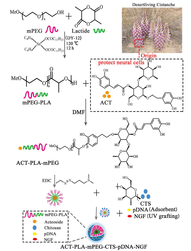
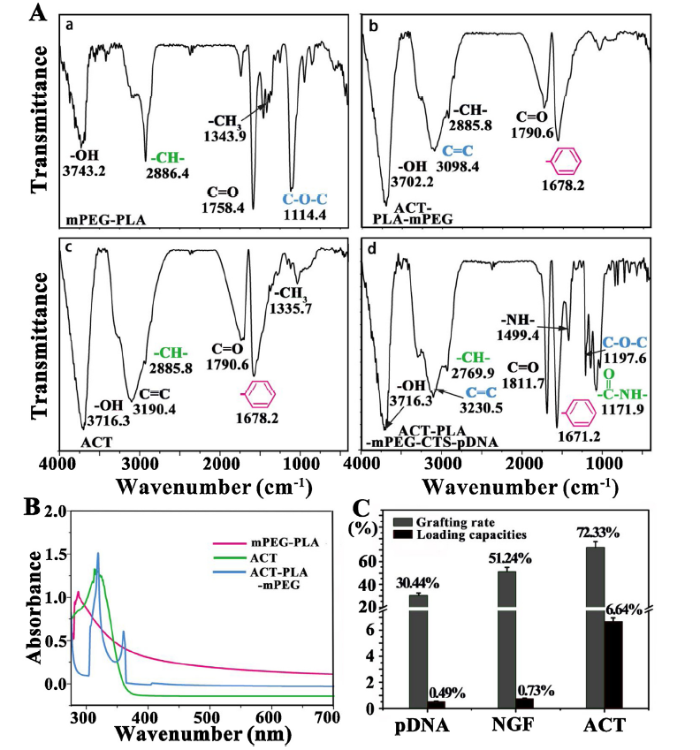
The stepwise process of synthesizing APPDN is depicted in Fig. 1. Fig. 2(A) shows the Fourier transform infrared spectroscopy (FITC) spectra of US, the mPEG-PLA spectra shows the absorption peaks at 1758.4 cm-1 and 1114.4 cm-1, which are the absorption peaks caused by the telescopic change of -OH and COC. It can be deduced that mPEG and PLA in the ACT-PLA-mPEG spectra were similar to those of the ACT-PLA-mPEG spectra at 3702.2 cm-1, 3098.4 cm-1, 1790.6 cm-1. And the ACT-PLA-mPEG spectra were similar to those of the ACT-PLA-mPEG-CTS, and 1678.2 cm-1 appeared four absorption peaks. -OH, C=C, C=O, benzene ring telescopic change, are ACT characteristic groups, that ACT successfully and mPEG in the DMF under the successful dehydration. The ACT-PLA-mPEG-CTS-pDNA has more spectral peaks than the characteristic spectrum of ACT, while the ACT-PLA-mPEG is aggregated and clustered with ACT-PLA-mPEG. The changes caused by 1494.4 cm-1 and 1197.6 cm-1, -NH-COC expansion were deduced respectively. Chitosan was initially formed by dehydration condensation to form amino acid acyl groups to synthesize ACT-PLA-mPEG-CTS-pDNA.
Fig. 1. Schematic illustration of the preparation of mPEG-PLA-ACT-CTS-pDNA-NGF (APPDN).
Fig. 2. (A) FITC spectra of mPEG-PLA, ACT-PLA-mPEG, ACT and ACT-PLA-mPEG-CTS-pDNA; (B) UV-vis spect of mPEG-PLA, ACT and ACT-PLA-mPEG; (C) The Grafting rate and Loading capacities of pDNA, NGF and ACT.
As shown in Fig. 2(B), the UV absorption peak of mPEG-PLA was approximately 280 nm (expressed as K absorption peak and was generated by groups containing conjugated double bonds). It was assumed that the carbonyl group in mPEG-PLA absorbed ultraviolet light. The ACT structure contained two benzene rings and several chromogenic groups. The higher the UV absorption, peak was closer to 336 nm. Compared to the ACT-PLA-mPEG UV absorption spectrum, the absorption shifted to the red, with the absorption peak appearing near the maximum absorption peak of ACT of around 360 nm. Hence, we speculated that ACT had successfully combined with the mPEG-PLA micelles. The grafting rate and drug loading capacities for pDNA, NGF and ACT were calculated using ultraviolet spectrophotometry (Fig. 2(C)). The grafting rates for pDNA, NGF and ACT were 30.44 %, 51.24% and 72.33%, respectively. The drug loading rates for pDNA, NGF and ACT were 0.49%, 0.73% and 6.64%, respectively.
The particle size and representative transmission electron microscopy images of the nano-drug-loading system are shown in Fig. 3(A). The morphology and particle size of the nano drug-loaded system were characterized using atomic force microscopy (Fig. 3(B)). The nanoparticles, prepared from a blend of mPEG-PLA and ACT-PLA-mPEG using the emulsion/solvent evaporation method had volume-based diameters of around 85 nm, and increased to around 100 nm after ACT was loaded. After surface modification with chitosan and electrostatic adsorption of pDNA and photografted NGF, the final particle size was about 160 nm. As shown in Fig. 3(C), Zeta potential of the mPEG-PLA formulation was about -20.33 mV, while that of ACT-PLA-mPEG was -16.57 mV. When CTS was used to modify the surface of micelles, the potential of the micelles decreased to equal that of CTS. After the nanoparticles adsorbed pDNA and NGF was grafted to its surface, the potential changed to positive. The APPDNs were then assessed for physical stability by suspending them in cell culture media and measuring their size using DLS (Fig. 3(D)). APPDN remained stable during testing and was not prone to condensation.
Fig. 3. (A) Size distribution histograms and TEM image of mPEG-PLA, ACT-PLA-mPEG, ACT-PLA-mPEG-CTS-pDNA and ACT-PLA-mPEG-CTS-pDNA-NGF; (B) AFM observation of ACT-PLA-mPEG-CTS-pDNA and ACT-PLA-mPEG-CTS-pDNA-NGF; (C) Zeta potential of mPEG-PLA, ACT-PLA-mPEG, ACT-PLA-mPEG-CTS-pDNA and ACT-PLA-mPEG-CTS-pDNA-NGF; (D) Variation of ACT-PLA-mPEG-CTS-pDNA-NGF particle size with time gradient in cell culture medium.
MPP+ + PC12 cells were used as the PD cell model [47] to investigate the neuroprotective effects of APPDNs. MPP+ + PC12 cell viability with or without the different nano-drugs were assessed using a standard MTT fluorescence assay, DAPI staining and annexin Y/PI double staining.
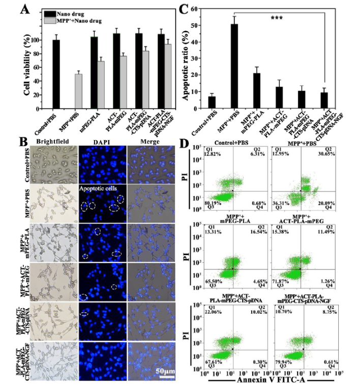
An MPP+ damage inducing in vitro cell model was established using PC12 cells. MTT assays were used to detect the repair effect of the nano drug delivery system on the MPP+ induced cell injury model (Fig. 4(A)). The cell viability of the MPP+ model group was significantly decreased to 51% compared to the blank control group, suggesting that the PD cell model had been successfully established. The different nanoparticle groups showed no toxicity in PC12 cells without the presence of MPP+ . More interestingly, all the experimental groups (i.e. MPP+ and nanoparticles) showed varying degrees of PC12 cell viability compared to the MPP+ model group. These results clearly demonstrated the neuroprotective effects of APPDNs at the cellular level.
Fig. 4. (A) Viabilities of PC12 cells 24 h after MPP+ treatment in the absence or presence of pre-incubated nano-drug with different groups were measured with MTT assays; (B) Bright field and confocal fluorescence images of PC12 cells treated with different groups. The nuclei of cells werestained with DAPI (blue); (C) Apoptosis rate of different groups were assessed by flow cytometry methods; (D) The PC12 cells apoptosis quantified by the flow cytometry at 24 h after various treated with different groups (PBS + PBS; MPP++PBS; MPP++ mPEG-PLA; MPP++mPEG-PLA-ACT; MPP++mPEG-PLA-ACT-CTS-pDNA; MPP++mPEG-PLA-ACT-pDNA-NGF) (Q1: dead cells; Q2: late apoptotic cells; Q3: live cells; Q4: early apoptotic cells)).
DAPI staining was then performed on PC12 cells treated with the different nano-drugs (Fig. 4(B)). Cell morphology was observed using light field microscopy and DAPI nuclear staining. The cell morphology of the control group was normal with an intact nucleus. In the MPP+ + PBS group, the nucleus was partially fragmented and the cell morphology was significantly altered. Treatment with mPEG-PLA resulted in improved cell and nuclear morphology, but was not very obvious. After ACT encapsulation, the cell recovery rate was similar to that of pure mPEG-PLA (lactic acid) micelles, however the nuclear morphology was significantly improved. After pDNA transplantation, cell morphology was significantly improved with a more complete nucleus. After APPDN treatment, there was an obvious cell recovery effect with nuclear integrity being restored. This demonstrated that the micellar complexes at different stages of synthesis had different therapeutic effects on the in vitro cell model. After pDNA and NGF grafting, the therapeutic effect was more significant.
The apoptosis rate of PC12 cells transfected with the above-mentioned formulations were next examined using flow cytometry (Fig. 4(C) and (D)). The cell survival rate in the control group was above 80%, while in the MPP++ PBS group, the apoptotic rate was more than 50%. This indicated that our Parkinson cell model was successfully established. After the cells were subjected to the different prodrug treatments, apoptosis rates for each group gradually decreased. After ACT encapsulation and grafting with pDNA and NGF, the therapeutic effect gradually increased. The apoptotic rate of APPDN, the synthesis end product of the nano-drug delivery system, decreased to less than 10%, and showed a remarkable neuroprotective effect.
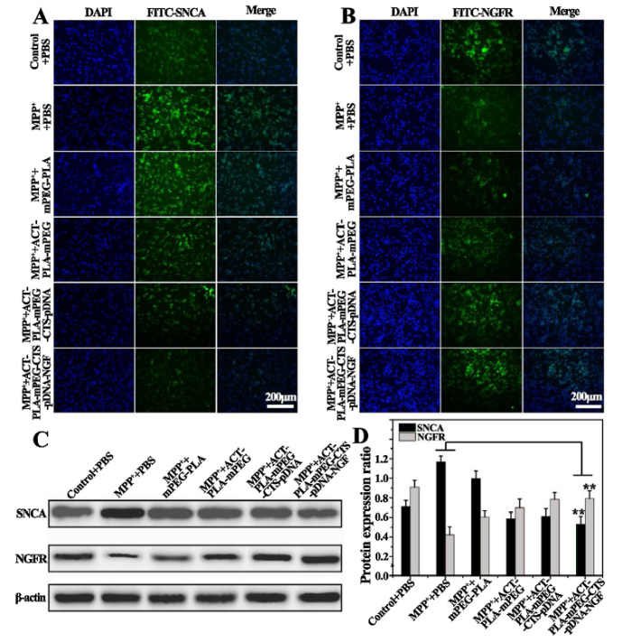
We next determined the effect of the nano-drug delivery system on the expression of α-synaptonuclear (SNCA) protein and nerve growth factor receptor (NGFR) in the PD cell model. We performed FITC fluorescence staining on PC12 cells from the different treatment groups. As shown in Fig. 5(A), the fluorescence intensity of α-syn was the highest in MPP+ + PBS group, which indicated that the PD cell model we constructed had the characteristics of abnormal α-syn aggregation. The fluorescence intensity of α-syn gradually decreased for each of the treatment groups, with the APPDN treatment group having similar levels as the control group. As shown in Fig. 5(B), the highest NGFR fluorescence intensity was in the control group and the lowest NGFR fluorescence intensity was in the MPP++ PBS group. NGFR fluorescence intensity increased gradually for the different treatment groups, with the APPDN treatment group having almost similar levels as the control group.
Fig. 5. (A, B) Confocal images of PC12 cells at 24 h after various treatments. Immunofluorescence staining was used to detect the nucleus (blue fluorescence) of DAPI, alpha synuclein (SNCA) and nerve growth factor receptor (NGFR) (green fluorescence) staining; (C, D) Expression of NGFR and SNCA after various treatment with different groups. The relative levels are plotted at a significance of p<0.05 indicated by *, 0.001<p<0.01 indicated by **, and p<0.001 indicated by ***, in comparison to the MPP++PBS group.
Next, we measured α-syn and NGFR protein levels by Western blotting. As shown in Fig. 5(C) and (D), α-syn expression levels in the control group was low, while α-syn levels in the MPP+ group was increased significantly. α-syn expression levels gradually decreased in all the treatment groups, with the APPDN group having the lowest levels, and was almost similar to that of the control group. NGFR protein levels in the PD model group were the lowest. NGFR protein levels gradually increased for the different treatment groups, with NGFR protein levels reaching the highest levels in the APPDN treatment group.
We then established the MPTP-induced PD mouse model [48] to evaluate the anti-PD effects of APPDNs. Female C57 mice at 7 weeks old were purchased from Southern Medical University and were used in all animal studies. Animals were divided into three groups, the control group (Saline + Saline), the model group (MPTP + Saline) and the treatment group (MPTP + APPDN).
By directly observing the experimental mice (Fig. 6(B)), i.e., tail, hair and other indicators, it was clear that mice in the treatment group were normal. None of the three groups showed significant weight changes during modeling and treatment (Fig. 6(C)).
Fig. 6. (A) Schematic representation of the open field system; (B) The photos of mice after various treatments (Saline + Saline; MPTP + Saline; MPTP + APPDN); (C) The body weight changes of mice during various treatments; (D-H) The behavior detection in mice after various treatments; (D, G) Open field test; (E) Pole-jump test; (H) Gait analysis Morris water maze (n = 5 biologically independent samples; two-way ANOVA with Tukey HSD test). The relative levels are plotted at a significance of p<0.05 indicated by *, 0.001<p<0.01 indicated by **, and p<0.001 indicated by ***, in comparison to the MPTP + saline group.
The efficacy of APPDN on reducing the symptoms of neurodegeneration and recovery of cognitive and locomotor activity was evaluated using the following behavioral tests: the pole test (Fig. 6(D)), gait analysis (Fig. 6(E)), open field test (Fig. 6(A) and (F) and (G)) and the water maze test (Fig. 6(H)), respectively. Compared to the control group, mice that were administered 8 d of continuous MPTP intraperitoneal injections showed significant PD symptoms, which included tremors, loss of balance, hypotonia, hollow back and hypokinesia. This suggested that the PD mouse model was successfully established.
The pole test (Fig. 6(D)) is a sensitive behavioral indicator for dopaminergic function [49]. MPTP-treated mice had increased time durations for “head down” and “climb down” while high-dosed APPDN treated mice had significantly reduced time durations compared to the model group. These results suggest that APPDN treatment could reduce α-synuclein accumulation and ameliorated behavioral impairment in subacute MPTP mouse models. For gait analysis (Fig. 6(E)), we measured step size for the different mouse groups and analyzed whether their gait was straight or not. As shown in Fig. 6(E), the stride length of PD mice was significantly shorter compared to mice in the normal control group. Gait analysis suggested that APPDN administration significantly improved dynamic gait function compared to MPTP-induced PD mice. This suggest that APPDNs ameliorates damaged motor function in PD mice.
Tests for anxiety-like behaviors in PD mice was performed using a simplified field experiment. Mice were observed for spatial and locomotor activity for five minutes. The open field system consists of a box that is separated into different areas and equipped with a camera. Fig. 6(A) depicts the open field system and contains different areas where mouse behaviors were recorded. As shown in Fig. 6(F) and (G), the three indicators that were measured included; the number of grid crossing, the time in the central grid and the number of standings and grooming. Compared to the control group, all three indicators in the model group were significantly reduced. However, after APPDN treatment, there was a significant improvement in all three indicators and were almost identical to the control group. In addition, a water maze study was performed to measure spatial exploration and memory ability. As shown in Fig. 6(H), mice in the treatment group had some improvements in spatial exploration, learning and memory ability compared to the model group. However, our results were confounded by the fact that mice in the control group had relatively long durations in spatial exploration, learning and memory ability. Additional studies would have to be performed to determine if APPDN treatment does in fact improve the three indicators mentioned above.
Tracer uptake in the brain area was observed in mice in the experimental group (MPTP + APPDN/CY5), with very minimal foci observed in control mice (MPTP + Saline/CY5) (Fig. 7(A1)). Semi-quantitative analysis was used to assess fluorescence intensity in the different groups (Fig. 7(A2)). Fluorescence intensity in the brains of mice in the MPTP + APPDN/CY5 group was much higher compared to mice in the MPTP + Saline/CY5 group. In addition, we observed that APPDN/CY5 reached the substantia nigra (a region implicated in the PD pathogenesis) after systemic administration (Fig. 7(B)). The above results demonstrated that APPDNs could pass through the BBB.
Fig. 7. (A1, A2) Representative images and head semi-quantitative analysis of APPDN/CY5 fluorescence imaging; (B) A representative picture of the retention of fluorescent labeled drugs in the substantia nigra at different times after intraperitoneal injection of APPDN/CY5 in mice. The nucleus is marked blue with DAPI and the APPDN is marked pink with CY5; (C, D) Immunofluorescence and alpha-syn immunohistochemical images of TH in substantia nigra of three groups (Saline + Saline; MPTP + Saline; MPTP + APPDN). The red circle indicates abnormal accumulation of alpha -syn cells, TH uses green fluorescent labeling; (E1, E2) Expression levels of TH and α-syn in the substantia nigra pars compacta of C57bl/6 mice and the quantitative results of ratio; (F) The images of H&E stained brain, lung, liver, spleen, kidney and heart after various treatments; (G-I) Serological and toxicology test of C57 mice, including the density of platelet, the density of white blood cell, and the density of red blood cell in different groups (Saline + Saline; MPTP + Saline; MPTP + APPDN). The relative levels are plotted at a significance of p<0.05 indicated by *, 0.001<p<0.01 indicated by **, and p<0.001 indicated by ***, in comparison to the MPTP + saline group.
Tyrosine hydroxylase (TH) is an enzyme responsible for catalyzing the conversion of L-tyrosine to dihydroxyphenylalanine (dopa), a precursor of dopamine. Alpha-syn (α-syn) is a soluble protein expressed in presynaptic and perinuclear areas of the central nervous system, which is closely related to the pathogenesis and related dysfunctions observed in Parkinson's disease. Immunohistochemistry, immunofluorescence staining and Western blot analysis for TH and α-syn expression levels were performed for the three experimental groups to evaluate the efficacy of APPDN. Immunofluorescence staining of TH in the substantia nigra is shown in Fig. 7(C) and immunohistochemical staining of α-syn in the dense part of the substantia nigra is shown in Fig. 7(D). In the model group, the number of TH positive dopaminergic neurons was reduced, while the number of α-syn positive dopaminergic neurons was increased. This demonstrated that MPTP + had an effect on TH and α-syn levels in the dense part of the substantia nigra. Damage to dopaminergic neurons results in over-expression of α-syn and down-regulation of TH. After APPDN administration, TH levels increased significantly while α-syn levels decreased significantly. These results suggests that APPDN could prevent dopamine neuron degeneration by up-regulating TH and down-regulating α-syn. Western blot results for TH and α-syn levels in brain tissues were consistent with IHC and IF results (Fig. 7(E)).
We assessed toxicity in vivo using HE staining of brain, lung, liver, spleen, kidney and heart tissues. As shown in Fig. 7(F), HE staining demonstrated that APPDN did not induce organ damage, and hence was safe for PD treatment. In addition, we assessed the toxicity of APPDN in PD mice by determining the number of blood platelets (PLT), white blood cells (WBCs), and red blood cells (RBCs). As shown in Fig. 7((G) and (H) and (I)), the number of WBCs increased to a certain extent in the model group (MPTP + Saline), but almost returned to normal levels in the treatment group (MPTP + APPDN). This suggested that there could have been a slight inflammatory reaction during the modeling process. Our results showed no significant differences compared to normal blood indices, indicating that there were almost no toxic effects in vivo.
Neurodegenerative disorders, including Parkinson’s disease (PD) and Alzheimer disease, represents a high social burden due to current treatment strategies being ineffective [50,51]. A major obstacle for the treatment of neurodegenerative disorders is the inability of therapeutic and diagnostic agents to cross the BBB and reach the affected brain regions [52]. The use of nanotechnology-based drug delivery approaches could overcome these challenges [53].
Chitosan is a novel nano drug carrier with targeting and sustained release, which has broad application prospects [36,54]. The nanoparticles prepared with chitosan as carrier have the advantages of protecting enzyme degradation, controlling release and improving bioavailability [55,56]. More importantly, chitosan can also enhance drug permeability by affecting the tight junction of the blood-brain barrier. These properties of chitosan contribute to the delivery of brain drugs, so chitosan nanoparticles are widely used as a brain-targeted drug delivery carrier. In addition, Current literature suggests that chitosan nanoparticles can bring about a convergence of drug and gene delivery [37,[57], [58], [59]]. So we synthesized a chitosan poly ethyleneglycol-poly lactic acid (PEG-PLA) nanoparticles conjugated with NGF, ACT and pDNA.
We have performed extensive studies using nanoparticles made of iron [60] and gold [61] to carry therapeutic payloads for the treatment of PD in animal models. Based on our previous studies, we established a nano-drug carrier system with low toxicity, ability to penetrate the BBB, was biodegradable and able to continuously release its payload in a controlled manner. Using this system we were successful in grafting ACT, pDNA and NGF to investigate its efficacy in PD mouse models.
FTIR detection, UV-vis absorption, particle size detection, TEM, AFM, and Zeta potential detection were used to demonstrate the successful synthesis of these nano-micelles and understand its morphology and chemical structure (Fig. 2, Fig. 3). The results demonstrated that the size of the nanoparticles was about 160 nm and had a positive charge. The size of the APPDN nanoparticles were regarded as favorable to pass the BBB and target the brain. These nanoparticles had enhanced permeability and retention (EPR) time and transversed the BBB through receptor-mediated transcytosis [62]. Considering the relative high levels of negative charge on the BBB, transcytosis occurs via electrostatic interactions between the positively charged moieties on the nanoparticles and the negatively charged membrane surface regions on brain endothelial cells [63].
The therapeutic effect of the APPDN nanoparticles in the PC12 cell model [47,64] was assessed using MTT and DAPI staining, flow cytometry, immunofluorescence and immunoblotting (Fig. 4, Fig. 5). Our results demonstrated that APPDN could effectively protect and repair cells after injury.
In addition, we established a PD in vivo model using C57 mice [48]. By evaluating behavioral traits (Fig. 6), we demonstrated that the model was successfully established and the nano-micelle could significantly improve PD symptoms in the model. Fluorescence imaging (Fig. 7(A) and (B)) studies demonstrated that APPDN could penetrate the BBB and play a therapeutic role. Western blot and immunohistochemical studies showed that APPDN could significantly improve PD symptoms in mice (Fig. 7(C) and (D) and (E)), and hence strongly supported its therapeutic effects. In addition, toxicity studies demonstrated that APPDN was safe after intraperitoneal injection (Fig. 7(F)-(I)).
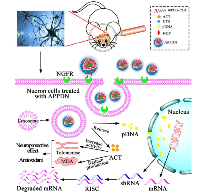
Polylactic acid crosslinked chitosan nanoparticles have been shown to improve drug delivery to the brain [65]. The polylactic acid crosslinked chitosan used in our APPDN improved the efficiency of the nanoparticles to pass through the blood-brain barrier, while mPEG directed the nanoparticles to dopaminergic neurons through electrostatic interactions with the brain extracelluar matrix (ECM). After the nanoparticles reached the dopaminergic neurons, the NGF that was grafted onto the surface of the nanoparticles interacted with the NGF-receptor on the surface of neurons, which then subsequently resulted in endocytosis of the nanoparticles into the cells. After nanoparticle entry, pDNA and ACT were released into the neurons to repair damaged dopaminergic neurons [[2], [3], [4], [5],66]. The synthetic nano-micelle inhibits the synthesis of α-synuclein to effectively reduce the formation of Lewy bodies through the neuroprotective effects of ACT (Fig. 8).
Fig. 8. Schematic illustration of the APPDN treatment process. The APPDNs nano- micelle complex is internalized by PC12 cells, and PDNA degrades mRNA through a series of actions, thus inhibiting SNCA expression. ACT inhibits cell senescence by increasing the activity of telomerase and reducing the production of malondialdehyde in the cytoplasm.
In summary, we developed a chitosan poly ethyleneglycol-poly lactic acid nanoparticle that was conjugated with NGF, ACT and pDNA. The size of the APPDN nanoparticle was about 160 nm. In addition, it had a positive charge, an optimal drug loading rate, low toxicity, was biologically degradable and penetrated the BBB easily. Our results demonstrated that APPDNs could significantly inhibit α-syn aggregation in vitro, which may be important for inhibiting Lewy body formation that was observed in our PD mouse model. In addition, APPDNs had significant neuroprotective effects on MPP+ lesioned PD cell models and in MPTP induced PD mouse models. For future studies we intend to further elucidate the mechanism of drug action and perform first-in-human studies.
This work was supported financially by the National Natural Science Foundation of China (Nos. 31370967 and 31170919), the Guangdong Province Universities and Colleges Pearl River Scholar Funded Scheme (2014), the Science and Technology Planning Project of Guangdong Province (No. 2015A020212033), the Science and Technology Project of Guangzhou (No. 201805010002), and the Innovation Project of Graduate School of South China Normal University (No. 2018LKXM019).

 WeChat
/
| 〈 |
|
〉 |


