Search for articles:
Chaoyi Yan , Chunyang Wu, Jie Xiong
Corresponding authors:
Received: 2019-07-10
Revised: 2019-08-26
Accepted: 2019-08-28
Online: 2020-04-01
Copyright: 2020 Editorial board of Journal of Materials Science & Technology Copyright reserved, Editorial board of Journal of Materials Science & Technology
About authors:
More
Abstract
High-performance electrocatalysts for water splitting are desired due to the urgent requirement of clean and sustainable hydrogen production. To reduce the energy barrier, herein, we adopt a facile in-situ surface modification strategy to develop a low-cost and efficient electrocatalyst for water splitting. The synthesized mulberry-like NiS/Ni nanoparticles exhibit excellent catalytic performance for water splitting. Small overpotentials of 301 and 161 mV are needed to drive the current density of 10 mA cm-2 accompanying with remarkably low Tafel slopes of 46 and 74 mV dec-1 for oxygen evolution reaction (OER) and hydrogen evolution reaction (HER), respectively. Meanwhile, a robust electrochemical stability is demonstrated. Further high-resolution X-ray photoelectron spectroscopy analyses reveal that the intrinsic HER activity improvement is attributed to the electron-enriched S on the strongly coupled NiS and Ni interface, which simultaneously facilitates the important electron transfer, consistent with the electrochemical impedance results. The post characterizations demonstrate that surface reconstructed oxyhydroxide contributes to the OER activity and NiS/Ni is an OER precatalyst. This structure construction with in-situ formation of active interface provides an effective way to design efficient electrocatalysts for energy conversion.
Keywords:
Electrocatalytic water splitting, as a promising technology for sustainable production of hydrogen fuel, has been the forefront research of energy conversion [[1], [2], [3]]. As a directly related part of electrolysis system, electrocatalyst plays the critical role in raising energy conversion efficiency [[3], [4], [5], [6], [7]]. More to the point, surface of catalyst is the location where the catalytic reaction occurs. The physical and chemical properties of surface directly determine whether the reaction takes place or not and the catalytic kinetics. Thus surface design is a key strategy to enhance the concerned catalytic performance [[8], [9], [10], [11], [12]].
To composite catalysts, the interface of hetero-material occupies a large proportion of the surface. The interface construction has been identified as an effective way to enhance catalytic activity of inactive materials [8,[13], [14], [15]]. Different from the internal defect engineering and surface crystallographic plane engineering [16,17], a rational interface can create new active sites by inducing unsaturated bondings, lattice distortion, and interface polarization. The generated electron redistribution tunes the initial density of states of surface atoms, which leads to optimized adsorptivity for hydrogen ions or water molecules and increased electron transfer ability [11,18]. In many cases, a new kind of active species grown on the surface of host material does not obtain a good electric contact. The interface resistance limits the reaction rate by impeding electron supply. Further annealing treatment is necessary to strength the binding [[19], [20], [21]]. Referring to this problem, the in-situ synthesis of active species on the surface of pristine host material can properly meet this requirement. The in-situ formation scheme well utilizes the atoms from the surface layer of host material and external solution or gas via the atomic diffusion reaction. Since there is no adsorption and deposition process of introduced second phase, the in-situ synthesis ensures a robust coupling between hetero-materials, which results in a strong electron interaction and a low charge transfer barrier on the interface [17,18,22]. This is beneficial for efficient electrocatalytic reactions.
In this work, we in-situ synthesized mulberry-like NiS/Ni nanoparticles on carbon cloth. The NiS/Ni composite exhibits excellent catalytic activity with remarkably low Tafel slopes of 46 mV dec-1 for oxygen evolution reaction (OER) and 74 mV dec-1 for hydrogen evolution reaction (HER) in alkaline solution. Small overpotentials of 301 and 161 mV are needed to afford the current density of 10 mA cm-2 for OER and HER, respectively. Further X-ray photoelectron spectroscopy analyses demonstrate that the superior activity of mulberry-like NiS/Ni composite is attributed to the strongly coupled interface between NiS and Ni, especially for HER. To OER, the evolved surface oxyhydroxide positively contributes to the OER activity. The internal sulfide and metal facilitate the charge transfer. Thus, the pristine NiS/Ni material acts as a functional OER precatalyst. This in-situ interface design is promising to be extended to novel catalyst development.
The Ni nanoparticles was prepared by a solvent thermal method. Firstly, Ni(OH)2/carbon cloth was prepared according to our previous work [23]. A piece of Ni(OH)2/carbon cloth was placed into a 50 mL Teflon-lined stainless autoclave with 30 mL ethanol. Then it was sealed and hold at 200 °C for 16 h in an electric oven. When the reaction over, the sample was taken out, washed with ethanol and dried.
The obtained Ni nanoparticles was annealed in a furnace with 10 mg S powder in the upper stream. The furnace temperature was rised to 290 °C and hold for 30-120 s. After a fast cooling process, the NiS/Ni sample was obtained.
Powder X-ray diffractometer (XRD, Rigaku Ultima IV) was used to analyze the crystal structure of synthesized sample. Scanning electron microscopy (SEM, FEI Nova NanoSEM 450) was used to characterize the morphology. Transition electron microscopy (TEM) analysis was performed on a FEI Tecnai G2 F20 microscope at 200 kV. The chemical composition and state investigations were carried out on a Kratos-Axis Supra X-Ray photoelectron spectroscopy (XPS) with a monochromatic Al Kα source.
All electrochemical measurements for HER and OER were performed in a three-electrode system on an electrochemical analyzer (CHI 660E or RST 5200 F) with a Ag/AgCl electrode as the reference electrode and a platinum foil as the counter electrode under room temperature in N2 or O2-saturated 1 M KOH. The exposed geometric area was 0.5 cm × 0.5 cm. The linear scanning voltammetry measurement was performed at a scan rate of 5 mV s-1. To evaluate the double layer capacitance (Cdl), cyclic voltammetry under different scan rates was tested in a non-faradic region of 0‒0.08 V for OER and from -0.82 to-0.92 V for HER, respectively. The electrochemical impedance spectroscopy (EIS) measurements were performed separately at 100 mV and 320 mV vs RHE for HER and OER from 100 k to 0.1 Hz with an AC voltage of 5 mV. All used potentials in the polarization curves were compensated with 95 % iR correction, which are given versus reversible hydrogen electrode (RHE) according to the equation:
ERHE=EAg/AgCl+0.197+0.059×pH(V)
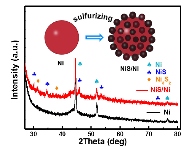
The interface design started with the basic preparation of Ni nanoparticles. It was obtained by a solvent thermal reduction method from nickel hydroxide. Then a facile thermal-treatment was performed along with a fast and mild surface sulfuration of the Ni nanoparticles. Fig. 1 and S1 show the X-ray diffraction (XRD) patterns of prepared samples. It is obvious that the diffraction peaks of NiS (JCPDS No. 02-1280) could be clearly identified after 60-150 s sulfuration reaction (depicted as NiS/Ni-time). The deeper sulfuration of Ni is limited by the annealing time, temperature and the amount of S powder. Thus the structure evolution process is controllable. Along with the grown of NiS, weak peaks from Ni3S2 impurity are detected by XRD. The inset scheme visually illustrates the structure evolution during sulfuration process with in situ formation of NiS nanocrystals.
Fig. 1. XRD patterns of Ni and NiS/Ni samples. Inset illustrates the surface structure evolution after sulfuration.
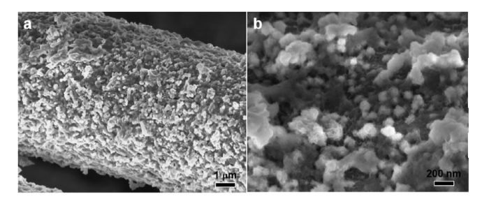
The concrete morphologies before and after surface sulfuration were investigated by scanning electron microscopy (SEM). Fig. 2(a) and (b) shows the prepared Ni nanoparticles covering the carbon fibers. The magnified image shows the particle size of 100-150 nm. The reaction time dependent structure evolution is shown in Fig. S2. With time going on, the surface NiS nanocrystals grow from about 5-40 nm. Deeper sulfuration can induce great change of the morphology. Within about 90-120 s sulfuration, mulberry-like composite nanoparticles in the size of about 100-200 nm are observed as shown in Fig. 2(c) and (d). A good coverage of target nanoparticles is maintained on the fibers. The in situ formed NiS nanocrystals in the size of 10-25 nm on the surface demonstrate the successful synthesis of heterostructure. This means that the accessible surface area is increased for electrochemical reaction. The heterostructure is also confirmed by the transition electron microscopy (TEM) analyses (Fig. 3 and S3). Fig. 3(a) shows the connected Ni precursor with smooth surface. The sulfurized nanoparticles are shown in Fig. 3(b) with formed surface nanocrystals less than 20 nm, consistent with above SEM results. Further high-resolution TEM image comfirms the crystal structure of synthesized sample in Fig. 3(c). The lattice distances of 0.207 nm and 0.299 nm correspond to the (111) plane of Ni and (100) plane of NiS, respectively. The coupled interface can be obviously observed. Additionally, the energy dispersive X-ray analysis element mapping verifies the local surface distribution of S on Ni nanoparticles, demonstrating the formation of surface NiS, in accordance with previous observations.
Fig. 2. Morphology characterizations: (a) and (b) SEM images of Ni nanoparticles, (c) and (d) SEM images of NiS/Ni samples under different magnifications, showing the in situ formed mulberry-like heterostructure. Inset shows the mulberry structure.
Fig. 3. Morphology and structure characterizations: (a) TEM image of Ni nanoparticles, (b) TEM image of NiS/Ni composite nanoparticles, (c) HRTEM image of NiS/Ni composite showing the interface coupling, (d) EDX elemental mapping images.
To verify whether such structure construction is beneficial to the electrolytic splitting of water, the electrochemical OER performance of NiS/Ni samples was firstly measured. The polarization curves in Fig. 4(a) demonstrate the effective structure modulation from surface sulfuration with optimized catalytic performance. The NiS/Ni-90 sample shows a 4.4 fold-current density improvement at 1.6 V after iR compensation compared to that of original Ni nanoparticles electrode. A current density of 10 mA cm-2 is achieved at a small overpotential of 301 mV. The reduced Tafel slope of 46 mV dec-1 of NiS/Ni-90, much lower than other sulfurized samples (63, 65 and 71 mV dec-1) and original Ni (69 mV dec-1), demonstrates the benefits of mild surface modulation (Fig. 4(b, c)). This suggests its high activity for oxygenic intermediate transformation and good charge transfer ability, better than most reported OER catalysts [13,19,[23], [24], [25], [26]]. The activity comparison in Fig. 4(c) clearly shows the structure-performance relationship under varied sulfuration degree, further supported by the decreased Rct value in the Nyquist plots by electrochemical impedance spectroscopy (EIS) measurement (Fig. S4). Obviously, NiS/Ni-90 exhibits the lowest overpotential of 338 mV to derive a current density of 30 mA cm-2. The larger oxidation peaks in polarization curves after deeper sulfuration are attributed to the easier surface structure evolution on NiS/Ni heterostructure. However, the deeper sulfuration also influences the integrity of internal Ni core, which in turn limits the charge transfer. It is known that the investigation of electrochemical active surface area (ECSA) is necessary. Typically, the calculation of double layer capacitances (Cdl) is performed to represent the ECSA, based on the varied-scan rate CV measurements. As shown in Fig. S5, NiS/Ni-90 electrode exhibits the largest Cdl value of 3.8 m F cm-2 in comparison with all other samples (2.0-2.8 m F cm-2), which suggests that the formation of surface heterostructure could induce a larger electrochemical surface area for OER. In addition, stability as an important criterion to evaluate electrocatalyst is necessary to be performed. The time-dependent current density (I‒t) measurement in Fig. 4(d) demonstrates the robust electrochemical stability of NiS/Ni-90 electrode during 60,000 s electrolysis at overpotential of 300 mV without clear performance attenuation (Fig. 4(d)). Inset shows the fast formation and release of O2 bubbles. The post-OER XRD analysis indicates the crystal structural degradation with obvious intensity attenuation of diffraction peaks (Fig. S6). It is known that sulfides and non-noble metals suffer inevitable surface oxidation during the OER process according to thermodynamic principles [27,28]. The irreversible and reversible structure phase transformations tend to occur at least on the surface layer, involving oxide, hydroxide and oxyhydroxide in the form of amorphous species or sheet-like nanostructure [[29], [30], [31], [32]].
Fig. 4. OER performance evaluation: (a) polarization curves of Ni and NiS/Ni nanoparticles at different sulfuration time, (b) corresponding Tafel slope, (c) activity comparison according to Tafel slope and potential at current density of 30 mA cm-2, (d) I‒t stability measurement at overpotential of 300 mV. Inset implies the fast formation and release of O2 bubbles.
To determine whether surface modulation of Ni is effective on facilitating the HER activity, we measured the HER performance of NiS/Ni samples. As shown in Fig. 5(a), the polarization curves of Ni after different sulfuration time exhibit different HER performance, better than pristine Ni nanoparticles electrode. The NiS/Ni-120 shows the best catalytic performance with a low overpotential of 162 mV to deliver the current density of 10 mA cm-2 and a small Tafel slope of 74 mV dec-1 after iR compensation. As shown in Fig. 5(b) and (c), the activity comparison shows a greatly decreased Tafel slope from 138 mV dec-1 to 74 mV dec-1 after 120 s sulfuration. Although the catalytic activity is strengthened, the electrocatalytic HER reaction still operates via the Volmer-Heyrovshy mechanism [33]. This means that supply of adsorptive proton plays as the primary rate determining step. The EIS analyses show a much smaller Rct value of NiS/Ni-120, about 35 Ω decrease compared to those of other samples, consistent with the polarization curve results (Fig. 5(d)). This indicates that a fast charge transfer path is built from the active interface to bottom electrode. Moreover, the ECSA is also evaluated by computing the Cdl values in Fig. 5(e) and S7, similar to the evaluation for OER. It is noting that the in-situ formed NiS nanocrystals increase the accessible electrochemical surface area of NiS/Ni with Cdl value of 8.2 m F cm-2 for NiS/Ni-120 compared to 3.7 m F cm-2 for Ni, also supported by aforesaid SEM and TEM analyses. Fig. 5(f) shows the stable electrolysis of NiS/Ni-120 for 67,000 s, which indicates that the NiS/Ni-120 electrode could serve as a robust HER catalyst. Inset shows the accumulation and fast release of H2 bubbles. It is noting that the post-HER NiS/Ni electrode also exhibits a slightly evolved surface with formed small nanosheets in Fig. S8. This is possibly attributed to the binding of hydroxyl after splitting of water molecules. On the other hand, it may involve the deposition of corrosive Ni ion with hydroxide ion.
Fig. 5. HER performance measurements: (a) polarization curves of Ni and NiS/Ni nanoparticles at different sulfuration time, (b) corresponding Tafel slope, (c) activity comparison according to Tafel slope and overpotential at current density of 10 mA cm-2, (d) Nyquist plots of Ni and NiS/Ni nanoparticles at different sulfuration time, (e) Cdl calculations for Ni and NiS/Ni nanoparticles at different sulfuration time, (f) I‒t stability measurement. Inset implies the fast formation and release of H2 bubbles.
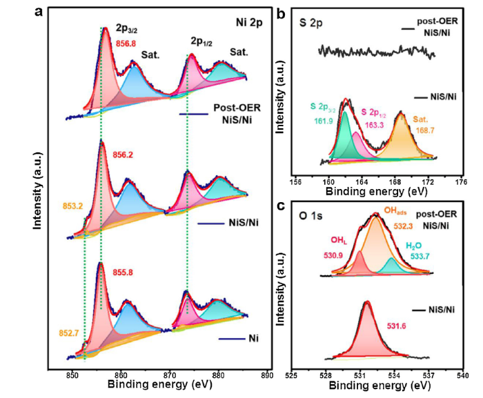
To verify the structure-performance relation of NiS/Ni catalyst, XPS was performed to investigate the surface chemical states of corresponding elements. The high-resolution Ni 2p spectra show a shift of 0.5 eV for Niδ+ (δ close to 0) towards higher binding energy (BE) after the sulfuration treatment in Fig. 6(a), which indicates the induced electron transfer from inner Ni core by strongly coupled NiS/Ni interface. On the other hand, the peak could also reveal the existence of Ni-Ni bonding in NiS domains. An electron-rich state after charge redistribution is beneficial to the hydrogen evolution reaction [14,34]. The S 2p spectra show a couple of peaks at 161.9 and 163.3 eV in Fig. 6(b), accompanying with a satellite peak at 168.7 eV. Compared to the S 2p regions in previous reports, the binding energy exhibits a shift toward lower binding energy [35]. This suggests the electron shift from Ni to S. It further supports the electron-rich state of S in NiS domains. As a result, the NiS/Ni shows an enhanced electrochemical performance for HER. However, to OER, we hypothesize that the interface structure facilitates the hydroxide formation as aforesaid. The coupled structure benefits the electron transfer from surface hydroxide layer to inner sulfide and metal nanoparticle during reaction. The evolved surface structure is further confirmed by the post-OER SEM investigations in Fig. 7, covering with obvious amorphous species and nanosheets. The nanoparticles are connected together by the formed species, which may benefit the internal carrier transfer. Besides, the post-OER XPS analyses show the disappearance of surface Niδ+ state and a BE shift of 0.6 eV toward higher BE of Ni in Fig. 6(a). This means the surface Ni exhibits a higher oxidative state after OER. Usually, the high-valence metal center is the adsorption site of oxygenic intermediates [36,37]. Meanwhile, the disappearance of S 2p peaks and the appearance of lattice OH (OHL, 530.9 eV) and adsorbed OH groups (OHads, 532.3 eV) in Fig. 6(b) and (c) demonstrate the surface transformation from NiS/Ni to Ni (oxy)hydroxide. Such results suggest that the activity of NiS/Ni electrode for OER is ascribed to the surface reconstructed (oxy)hydroxide. NiS/Ni plays the roles of precatalyst, electron conductor and electronic structure modulator. It shows good consistence with previous reports towards transition-metal derived OER electrocatalysts [38]. Besides, as recently reported, Fe impurity from the electrolyte may influence the catalytic activity of OER catalysts [39]. Here, we further performed high-resolution XPS to detect the possible Fe impurity in the surface Ni (oxy)hydroxide layer with Mg Kα source. As shown in Fig. S9, a weak peak of Fe 2p3/2 at 711.7 eV is detected with a low content about 0.44 at.%. It is known that the existence of Fe in the lattice of Ni (oxy)hydroxide is responsible for the high OER performance [39]. Thus we propose that the reconstructed Fe-doped Ni (oxy)hydroxide on the surface of NiS/Ni contributes to the high OER activity.
Fig. 6. XPS characterizations before and after OER: (a) Ni 2p spectra of Ni, NiS/Ni-90 and post-OER NiS/Ni-90 electrodes, (b) S 2p spectra of initial NiS/Ni-90 and post-OER NiS/Ni-90 electrodes, (c) O 1s spectra of initial NiS/Ni-90 and post-OER NiS/Ni-90 electrodes.
Fig. 7. (a) and (b) SEM images of NiS/Ni-90 electrode after OER stability measurement, showing formed amorphous species and nanosheets on the surface.
In summary, a highly-efficient electrocatalyst for water splitting is designed by a surface modification strategy. Via controllably tailor the sulfuration degree, the constructed mulberry-like NiS/Ni composite catalysts exhibit excellent catalytic activity for both HER and OER, with low Tafel slope of 74 mV dec-1 and 46 mV dec-1, respectively, accompanying with robust durability. Benefiting from the strongly coupled NiS/Ni interface, the enriched electrons on in-situ formed NiS domains contribute to the HER performance improvement. Moreover, the NiS/Ni interface facilitates the surface oxidation with higher oxidative state of Ni for catalyzing oxygen evolution. The identified Fe impurity on the surface is positively beneficial to the activity improvement. This facile approach for interface structure design is promising to be applied to other reactions in energy conversion field beyond water splitting.
This work was financailly supported by the National Natural Science Foundation of China (Nos. 51722204 and 51802145), the National Key Basic Research Program of China (No. 2014CB931702), the Sichuan Science and Technology Program (Nos. 2018RZ0082 and 2019JDRC0070), the Fundamental Research Fund for the Central Universities (No. A03018023801053) and the Open Project of Jiangsu Key Laboratory for Carbon-Based Functional Materials Devices at Soochow University (No. KJS1807).
Supplementary material related to this article can be found, inthe online version, at doi:https://doi.org/10.1016/j.jmst.2019.08.042.

 WeChat
/
| 〈 |
|
〉 |


