Search for articles:
Huaqiang Zhuang , Wentao Xu
Corresponding authors:
Received: 2018-12-19
Revised: 2019-05-6
Accepted: 2019-05-10
Online: 2019-10-05
Copyright: 2019 Editorial board of Journal of Materials Science & Technology Copyright reserved, Editorial board of Journal of Materials Science & Technology
More
Abstract
The one-dimensional ZnWO4@SnWO4 photocatalyst with a core-shell heterostructure was successfully constructed by a simple two-step method. It is interesting to note that ZnWO4@SnWO4 composite photocatalyst owns a higher photocatalytic activity for RhB degradation under visible light irradiation. The introduction of SnWO4 shell layer, which forms a clear heterogenous interface between ZnWO4 and SnWO4, increases the photo-absorption efficiency of ZnWO4 nanorods. In addition, its band-edge absorption evidently shifts toward the visible region. Based on the photoelectrochemical (PEC) and electron spin resonance (ESR) measurements, it is found that the photocatalytic activity was attributed to the efficient separation and transfer of photo-generated charge carriers. Hence, they can produced more hydroxyl radical (•OH) as the main active species in the photocatalytic reaction process.
Keywords:
Currently, the abundant discharge of textile industrial effluent is becoming a huge challenge to public health and environment with the development of the economy and industry [1,2]. Hence, an efficient pollution-free processing technology to remove the toxic contaminants from water waste is highly imperative and desired [[3], [4], [5]]. Semiconductor-based photocatalysis is regarded as one of the most promising technologies [[6], [7], [8]]. Since 2006, Zhu et al. [9] first found that ZnWO4 owned the high photocatalytic activity for the degradation of RhB, ZnWO4-based photocatalysts began to be used for the degradation of organic pollutants. Due to its unique physicochemical properties, ZnWO4 can be used as scintillating materials, optical fibers, gas sensors, photocatalysts, etc. [10]. However, ZnWO4 is a wide band-gap semiconductor and has lower charge transfer efficiency, which severely limits its practical applications [11]. Hence, many strategies such as doping, semiconductor compositing, and sensitization have been carried out to overcome the existing problems. Recently, Chen et al. [12] reported that ZnWO4-x sample with oxygen vacancies can degrade 91% and 78% tetracycline under the irradiation of UV and ultraviolet-visible-near infrared (UV-Vis-NIR) light, respectively. Ran et al. [13] found that Si-doped ZnWO4@ZnO nanocapsules with open-shaped structures displayed the high effective photocatalytic activity for degradation of RhB under visible light irradiation. In addition, GO/ZnWO4 [14], ZnWO4/ZnFe2O4 [10] and ZnWO4/Cu2O [15] composites also showed the enhanced photocatalytic activity. Therefore, it can be concluded safely that the semiconductor heterostructure by coupling a suitable semiconductor is a good way to improve photocatalytic performance
Stannous tungstate (SnWO4) is a promising candidate as a visible-light-sensitive photocatalyst for its desired band-gap and unique electronic catalytic property [16,17]. It is well known that SnWO4 has two crystalline phases (including α-phase and β-phase), and both of them can be appropriate for the photocatalysis [16,18]. Compared with the other photosensitizers, the SnWO4 material displays several distinct advantages: (1) SnWO4 material is a narrow band gap semiconductor and has appropriate band-gap position; (2) It can be easily prepared by in-situ synthesis method; (3) SnWO4 and ZnWO4 materials are tungstate with the same ABO4 structure. Thus, it is very intriguing to explore the performance of photocatalyst that the SnWO4 material is introduced as photosensitizer on surface of ZnWO4 nanocrystal to fabricate the heterostructure.
In this work, the ZnWO4@SnWO4 heterostructure composite with a core-shell structure was successfully fabricated by a simple two-step method. The as-prepared ZnWO4@SnWO4 material exhibits an excellent photocatalytic activity for RhB degradation under visible light irradiation. Furthermore, the detailed structure and properties of photocatalysts were also analyzed by a series of characterizations. Thereinto, the uniform core-shell structure and a clear heterogenous interface between ZnWO4 and SnWO4 could be observed by SEM and TEM measurements. It was found that the incorporation of SnWO4 shell layer could not only enhance photo-absorption efficiency of ZnWO4 nanorods, but also drastically improve its photocatalytic performance under visible light irradiation. Meanwhile, PEC and ESR measurements were also used for the photocatalytic reaction mechanism, suggesting that the enhanced photocatalytic activity was attributed to the more efficient separation and transfer of photo-generated charge carriers, and they could produce more hydroxyl radical (•OH) as the main active species in photocatalytic reaction process.
ZnWO4 nanorods were prepared by a simple hydrothermal synthesis [19]. In a typical process, 3 mmol Zn(NO3)2•6H2O and 3 mmol Na2WO4•2H2O were dissolved together in 28 mL deionized water under 30 min vigorous stirring. Then, the mixed solution was transferred into a stainless-steel autoclave with a Teflon cup of 50 mL inner volume. Subsequently, the pH value of mixed solution was adjusted to 7 using 0.5 mol/L NaOH, and the mixture was stirred for 1 h at room temperature. After being sealed, the autoclaves were heated in a convection oven at 180 °C for 48 h. Finally, the white precipitate was obtained and washed with distilled water and ethanol by centrifugation, and then dried in an oven at 60 °C for 12 h.
ZnWO4@SnWO4 nanorod composite was prepared by an in-situ synthetic method. 2 mmol ZnWO4 nanorods and 80 mL of distilled water were added into a 150 ml three-necked flask, and the dispersion was refluxed at 120 °C. Then, the pH of mixed solution was adjusted to 5 using 0.1 mol/L HCl. Subsequently, 0.2 mmol SnCl2•2H2O and 1 mL of 0.2 mol/L Na2WO4 solution were added into the mixed solution each time at an interval of 3 h for several times. After being added, the mixed solution was continuously refluxed at 120 °C for 3 h. The obtained yellow sample was washed with distilled water and ethanol by centrifugation, and then dried in an oven at 60 °C for 12 h. SnWO4 sample using the above synthetic method was prepared, except that ZnWO4 nanorods didn’t be added. The sample denoted as ZnWO4/SnWO4 was prepared by the mechanical mixing of SnWO4 with ZnWO4, and the mass ration of ZnWO4 and SnWO4 is equal for ZnWO4/SnWO4 and ZnWO4@SnWO4 samples.
X-ray diffraction (XRD) plots were measured on a Bruker D8 Advance X-ray diffractometer with CuKα radiation (1.5406 Å). The Hitachi New Generation SU8010 field emission scanning electron microscope (SEM) was employed to obtain the SEM images. High resolution transmission electron microscopy (HRTEM) images were obtained by a JEOL model JEM 2010 EX instrument at an accelerating voltage of 200 kV. X-ray photoelectron spectroscopy (XPS) were carried out on a VGESCALAB 250 XPS system with a monochromatized AlKα X-ray sources (15 kV 200 W 500 μm pass energy = 20 eV). All binding energies were referenced to the C 1s peak at 284.6 eV of surface adventitious carbon. UV-vis DRS spectra were taken on a Varian Cary 500 Scan UV-vis-NIR spectrometer with BaSO4 as the reference. Electron spin resonance (ESR) spectra were measured using a Bruker model A300 spectrometer with a 500 W Xe-arc lamp equipped with an UV-cutoff (≥ 420 nm) as visible light source. Typically, the water or methanol solution of DMPO was added to 5 mg samples, and the mixture was sampled using a capillary tube and placed in an ESR tube for the experiment.
Photoelectrochemical tests were performed using the electrochemical workstation (CHI 660E). Photocurrent was measured using the conventional three-electrode electrochemical cell with a working electrode, a platinum foil counter electrode and an Ag/AgCl electrode as reference electrode. The working electrode was fabricated by a special way to deposit a certain amount of catalyst onto the fluorine-doped tin oxide (FTO) glass. The working electrode was fabricated on the FTO glass: 10 mg of sample was mixed with 0.5 ml ethanol with continuous stirring for 3 h to get slurry. Then, 20 μL of slurry was spreading onto FTO glass, which side part was previously protected by Scotch tape, and the electrode was dried at room temperature for 24 h. In this experiment, the three electrodes were immersed in a mixed solution with a 0.2 mol/L Na2SO4 aqueous solution, and the working electrode was irradiated by visible light (≥ 420 nm).
The photocatalytic performance of the as-prepared samples was evaluated by the degradation of RhB in aqueous solution. A 300 W Xe lamp with a 420 nm cutoff filter was used as the light source to ensure the irradiation with visible light only. Typically, 80 mg of photocatalyst was added into 80 mL of 10 mg/L RhB aqueous solution in a container. Prior to irradiation, the suspensions were magnetically stirred in dark for 60 min to ensure the establishment of an adsorption/desorption equilibrium between the photocatalyst and RhB. At given irradiation time intervals, 4 mL of suspension was collected and centrifuged to remove the photocatalyst particles, then the residual RhB solution was analyzed by monitoring variations at the wavelength of maximal absorption (554 nm) in the UV-vis spectra.
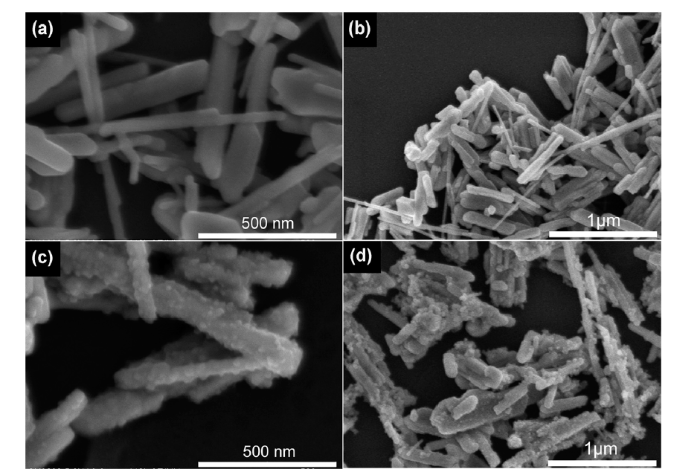
Fig. 1 shows SEM images of ZnWO4 and ZnWO4@SnWO4 nanorods. For the nude ZnWO4 sample, it can be observed that the ZnWO4 nanorods were in the range of 500-1000 nm length with a smooth surface, as shown in Fig. 1(a) and (b). Intriguingly, an uneven SnWO4 shell layer is completely covered on the smooth surface of ZnWO4 nanorods, as displayed in Fig. 1(c). The SnWO4 nanoparticles are uniformly dispersed on the surface of ZnWO4, indicating that the heterostructure between ZnWO4 and SnWO4 can be fabricated. In addition, Fig. 1(d) shows that the great majority of ZnWO4 nanorods can be uniformly coated with SnWO4 nanoparticles by the in-situ synthetic method.
Fig. 1. SEM images of (a, b) ZnWO4 nanorods and (c, d) ZnWO4@SnWO4 heterostructure.
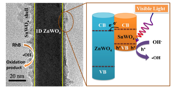
The ZnWO4 and ZnWO4@SnWO4 samples were further examined by transmission electron microscopy (TEM). Fig. 2(a) shows the smooth and well-proportioned surface for the pure ZnWO4 nanorods, which agrees well with the SEM results. Meanwhile, the core-shell structure of ZnWO4@SnWO4 sample is clearly display in Fig. 2(b) and (c). It is can be found that the SnWO4 nanoparticles are fully coated on the entire surface of ZnWO4 nanorods, which presents the ideal shell layer. Moreover, the core-shell structure was further investigated by HRTEM, as shown in Fig. 2(d). The distinct interface between ZnWO4 and SnWO4 can be clearly observed, suggesting that the heterostructure was successfully fabricated. Additionally, the interplanar spacing of 0.373 nm can be attributed to the (011) crystal plane of ZnWO4, which corresponds to the previous report [20]. However, the lattice fringe of SnWO4 shell layer can’t be observed in HRTEM image, implying that the SnWO4 shell layer is amorphous.
Fig. 2. TEM images of (a) ZnWO4 nanorods and (b, c) ZnWO4@SnWO4 heterostructure, (d) HRTEM image of ZnWO4@SnWO4 heterostructure and (e) energy dispersive spectroscopy mapping images of ZnWO4@SnWO4 heterostructure.
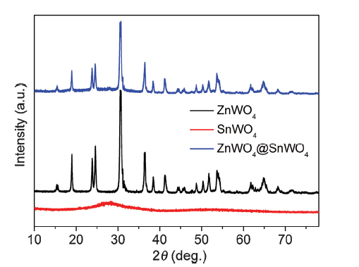
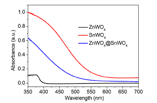
Fig. 3 shows the XRD pattern of all as-prepared samples. Apparently, SnWO4 sample displays an amorphous phase with a weak and broad characteristic peak, which agrees with the result of HRTEM measurement. Meanwhile, the characteristic diffraction peaks of ZnWO4 sample can be well assigned to pure monoclinic ZnWO4 (JCPDS card no. 73-0554) [21]. Interestingly, the intensity of diffraction peaks of ZnWO4 sample becomes weaker after the SnWO4 shell layer coated, suggesting that SnWO4 nanoparticles are deposited on the surface of ZnWO4 nanorods. This is in line with the results of SEM and TEM measurements. Ultraviolet-visible diffuse reflectance absorption spectra (DRS) were employed to investigate the optical absorption properties of SnWO4, ZnWO4 and ZnWO4@SnWO4 samples, as shown in Fig. 4. It can be seen that the SnWO4 sample displays remarkable absorption capability in the visible light region, and the absorption band edge of ZnWO4 sample is observed in the UV region [22]. As expected, the absorption band edge of ZnWO4@SnWO4 sample appears the red-shift to visible light region, which can be due to the introduction of SnWO4 shell layer.
Fig. 3. XRD patterns of ZnWO4, SnWO4 and ZnWO4@SnWO4 samples.
Fig. 4. UV-vis DRS spectra of ZnWO4, SnWO4 and ZnWO4@SnWO4 samples.
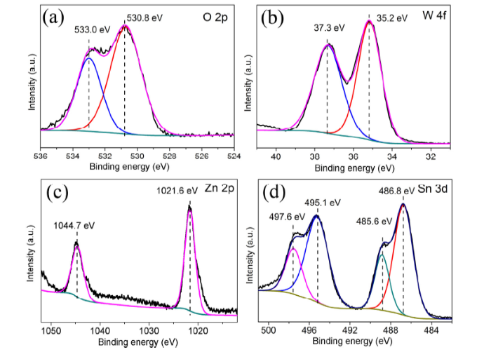
XPS was employed to investigate the chemical and binding state of SnWO4 nanoparticles on the surface of ZnWO4 nanorods, as shown in Fig. 5. Fig. 5(a)-(d) displays the high resolution XPS spectra for O 2p, W 4f, Zn 2p and Sn 3d, respectively. Fig. 5(a) shows the binding energy of O 1s at 530.8 eV and 533.0 eV. The lower binding energy peak at 530 eV can be attributed to the metal bound oxide component (O2-) of [WO4]2-, and the peak at 533.0 eV is assigned to the surface adsorbed or structural water molecules [[23], [24], [25]]. The XPS spectrum of W 4f shows two characteristic peaks at around 35.2 eV for W 4f7/2 and 37.3 eV for W4f5/2, respectively, suggesting that W exists as W6+ in ZnWO4@SnWO4 nanorods [24,26]. Meanwhile, the binding energy of Zn 2p also can be observed at 1021.6 eV and 1044.7 eV, which is attributed to Zn2+ in ZnWO4 nanorods [23]. In addition, the XPS spectrum of Sn 3d can be clearly observed, which further demonstrates the existence of amorphous SnWO4 nanoparticles. However, the Sn 3d XPS spectrum of ZnWO4@SnWO4 nanorods displays two core level peaks at ca. 486.8 eV and 488.9 eV, which is assigned to Sn2+ in SnWO4 and Sn4+ species, respectively [24,26]. It suggests that a small parts of Sn2+ ion is oxidized into Sn4+ species in the prepared process. Combining with the result of XRD pattern, the SnO, SnO2 or Sn3O4 samples did not be detected in the SnWO4 sample or ZnWO4@SnWO4 nanorods. Those results demonstrate that the Sn4+ species may be Sn4+ ion, which is adsorbed on the surface of ZnWO4@SnWO4 nanorods, so it can be detected by XPS measurement.
Fig. 5. High resolution XPS spectra of (a) O 2p, (b) W 4f, (c) Zn 2p and (d) Sn 3d for ZnWO4@SnWO4 heterostructure.
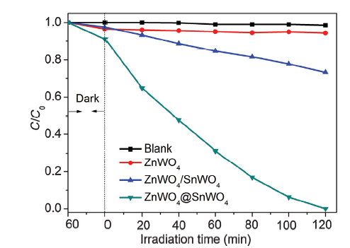
Photocatalytic activity of the Blank, ZnWO4, ZnWO4/SnWO4 and ZnWO4@SnWO4 samples was evaluated for RhB degradation under visible light irradiation (λ ≥ 420 nm), as shown in Fig. 6. The blank experiment for photolysis of RhB was carried out. Apparently, the decomposition of RhB is inappreciable in the absence of photocatalyst, indicating that the RhB is stable under the experiment condition. Similarly, ZnWO4 sample also displays the weak photocatalytic activity after 120 min of irradiation, because it is a wide-band-gap semiconductor. Interestingly, when ZnWO4 and SnWO4 semiconductors were coupled together, it presented an excellent photocatalytic activity for the degradation of RhB under visible light irradiation. It can be seen that the removal rate of RhB reached to 100% within 2 h. It suggests that the SnWO4 shell layer acting as a visible light photocatalyst plays an important role in the photocatalytic reaction process. In order to testify the advantage of the ZnWO4@SnWO4 core-shell structure, the ZnWO4/SnWO4 sample used as the reference samples was prepared and evaluated for RhB degradation. Obviously, the ZnWO4/SnWO4 sample with the mechanical mixing displayed a lower photocatalytic performance, suggesting that the enhanced performance is dependant on their type of unique and excellent structure.
Fig. 6. Photocatalytic activity of RhB degradation for Blank, ZnWO4, ZnWO4/SnWO4 and ZnWO4@SnWO4 samples under visible light irradiation.
In order to explore the mechanism for enhanced photocatalytic performance of ZnWO4@SnWO4 sample, the photoelectrochemical measurement was carried out, as shown in Fig. S1 in supporting information. Generally speaking, the photo-generated current intensity can indirectly reflect separation efficient of photo-induced electrons and holes [27]. Evidently, the photocurrent density of ZnWO4@SnWO4 sample is much higher than that of ZnWO4/SnWO4 and ZnWO4 sample. The increased photocurrent response suggests that the separation and migration efficiency of photogenerated charge carries are greatly improved, which is beneficial to the enhancement of photocatalytic activity.
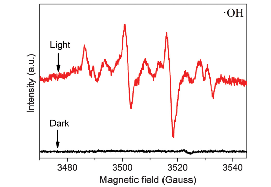
The specific active species of ZnWO4@SnWO4 sample was further explored by electron spin resonance (ESR) measurement in photocatalytic reaction process, as shown in Fig. 7. Obviously, the four characteristic peaks of DMPO-•OH adducts can be clearly observed, indicating that the OH- can be oxidized into •OH by the photogenerated holes in the valence band of ZnWO4@SnWO4 sample. On comparison, there is no signal of DMPO-•OH adducts under dark test condition. It indicates that the hydroxy radical (•OH) are indeed produced in the reaction system of ZnWO4@SnWO4 photocatalyst in aqueous solution under visible light irradiation. Simultaneously, in order to verify whether the superoxide radical (•O2-) can be generated in the photocatalytic process, the ESR measurement of ZnWO4@SnWO4 photocatalyst can be carried out in DMPO methanol solution under visible light irradiation, as displayed in Fig. S2. However, there is no signal of DMPO-•OH adducts in DMPO methanol dispersion. In conclusion, it demonstrates that the •OH is main active species in the photocatalytic process.
Fig. 7. ESR signal of DMPO-•OH spin adducts for ZnWO4@SnWO4 heterostructure under visible light irradiation.
Based on the above results, the reaction mechanism for enhanced photocatalytic activity can be proposed in Scheme 1. The SnWO4 sample is stimulated to produce the photo-induced electrons and holes under visible light irradiation. Because the flat band potential of SnWO4 sample is higher than that of ZnWO4 sample (Fig. S3), the photogenerated electrons on conduction band (CB) of SnWO4 can be quickly transferred to conduction band (CB) of ZnWO4. And the holes on valence band (VB) of SnWO4 can react with OH- and produce more hydroxyl radical (•OH) as main active species. Finally, the organic pollutant was completely degraded by the hydroxyl radical (•OH).
Scheme 1.. Proposed mechanism of ZnWO4@SnWO4 heterostructure for enhanced photocatalytic activity.
In this work, we designed and fabricated the one-dimensional ZnWO4@SnWO4 heterostructure composite with a core-shell structure, which displayed an excellent visible-light photocatalytic activity for RhB degradation. The introduction of SnWO4 shell layer can not only enhance the absorption and utilization of visible light for ZnWO4 nanorods, but also promote charge transfer and separation efficiency so that to improve its photocatalytic activity. The enhanced performance can be attributed to the matched level structure and the intimately contacted interface. In addition, the hydroxyl radical (•OH) can be regarded as the main active species in the photocatalytic reaction process. This work exhibits a novel ZnWO4@SnWO4 core-shell heterostructure for efficient visible light photocatalysis, so we can believe that it can be further applied to the designing of other semiconductor photocatalysts for improving their photocatalytic performance.

 WeChat
/
| 〈 |
|
〉 |
