Journal of Materials Science & Technology, 2016, 32(9): 840-844
doi: 10.1016/j.jmst.2016.04.009
Novel Gelatin-based Nano-gels with Coordination-induced Drug Loading for Intracellular Delivery
Changjiang Fan1,2, Dong-An Wang1,*,
1 Division of Bioengineering, School of Chemical and Biomedical Engineering, Nanyang Technological University, 70 Nanyang Drive, N1.3-B2-13, Singapore 637457, Singapore
2 Institute for Translational Medicine, College of Medicine, Qingdao University, Qingdao 266021, China
In this study, we develop the gelatin-dopamine (Gel-Dopa) nano-gels (GDNGs) and explore their potential as drug delivery vehicles. The Gel-Dopa precursor is synthesized using EDC/NHS coupling reaction, in which the catechols can coordinate with transition metal ions such as Fe3+. These novel GDNGs exhibit excellent cytocompatibility. The model drug, doxorubicin (Dox), is readily conjugated into catechol of GDNGs by the coordination cross-link of Fe3+ ion. The morphology and size distribution of the nano-gels are characterized via field emission scanning electron microscopy and particle size analyzer, respectively. The GDNGs loaded with Dox (GDNGs-Dox) is capable of efficiently penetrating cell membrane and enter the HeLa cells. The endocytosed GDNGs-Dox release Dox molecules and subsequently kill the tumor cells.
Various nano-particles have been prepared from numerous natural or synthetic materials and widely employed as drug delivery vehicles, aiming to enhance therapeutic effects and minimize side effects of drug formulation[1], [2] and [3]. Gelatin is derived from animal collagen through acid or alkaline hydrolysis; it is biocompatible, and relatively cheap as well as readily available[4], [5] and [6]. Like collagen, gelatin bears some cell recognized moieties (such as RGD sequence); the multiple functional groups (e.g. -COOH, -NH2) endow gelatin with the opportunity to be modified using various targeting ligands[5]. Besides, gelatin exhibits low anti-genicity and good solubility due to its denatured property[6]. Gelatin has been considered “generally safe” material by the United States FDA[7].
Gelatin nano-gels have been extensively used for the delivery of anticancer drugs, such as doxorubicin (Dox)[8]. Gelatin nano-gels can be easily prepared by many methods (such as desolvation, emulsification, coacervation, and nanoprecipitation); they are generally cross-linked with glutaraldehyde(GA)[9], genipin[10], or carbodiimide/N-hydroxysuccinimide[11]. The special characterizations of gelatin (e.g. low cost, cytotoxocity and anti-genicity, good solubility) offer many advantages for the applications of gelatin nano-gels in drug delivery. Gelatin nano-gels can be effectively internalized and localized into cells, indicating gelatin nano-gels have the ability to overcome cellular barriers and serve as efficient intracellular drug delivery vehicles[8], [12] and [13]. More importantly, gelatin nano-gels possess passive targeting capability via the enhanced permeability and retention (EPR) effect and then accumulate in tumors for plenty of time, which ensures the release, concentration, and work of the loaded drugs in situ[8] and [14].
Various strategies have been explored to load drugs into gelatin nano-gels, such as physical interactions (e.g. hydrogen bonds, hydrophobic interactions)[15] and [16], electrostatic interactions[17], and covalent conjugation[9]. Recently, coordination bond has emerged as an intriguing method to prepare nano-complexes for biomedical applications[18], [19], [20] and [21], and which may propose a new avenue to assemble drug into polymer nano-particles. Compared with the common methods for drugs' loading, complexation-driven drug installing exhibits intrinsic stability and pH-responsiveness based on the selection of different transition metals[18] and [22]. Besides, the complexation-based drug loading is facile and versatile[18].
In this study, we develop novel gelatin-dopamine (Gel-Dopa) nano-gels (GDNGs) as drug delivery vehicles. The design rationale of GDNGs is to achieve coordination complexation based drug loading into gelatin-based nano-gels and drug release after internalization into tumor cells (Fig. 1). Gel-Dopa conjugate is prepared by grafting dopamine onto gelatin backbone, and acts as the precursor to fabricate GDNGs. The GDNGs are formed by an established desolvation process and accompanying GA cross-link (Fig. 1(B)-a). The model drug Dox is simply loaded into GDNGs through the coordination interactions between Fe3+ ions and ligands (including catechols in GDNGs and phenolic groups in Dox) (Fig. 1(B)-b). The morphology and size distribution are studied by FESEM and a particle size analyzer, respectively. Cellular uptake of Dox-loaden GDNGs (GDNGs-Dox) is observed under a fluorescence microscope. The in vitro cytotoxicities of GDNGs and GDNGs-Dox are evaluated in detail.
Fig. 1.
(A) Synthesis scheme of Gel-Dopa precursor via EDC/NHS chemistry. (B) Schematic fabrication process of GDNGs-Dox from Gel-Dopa precursor: (a) GDNGs are first synthesized via a desolvation method; subsequently, (b) the GDNGs suspension is stirred with Dox in the presence of FeCl3, achieving GDNGs-Dox. (C) Schematic illustration of targeting and endocytic process of GDNGs-Dox by a tumor cell.
2. Materials and Methods
2.1. Synthesis of gel-dopa
The dopamine is grafted onto gelatin by ethyl-dimethyl-aminopropylcarbodiimide (EDC) and N-hydroxy-succinimide (NHS) coupling chemistry[23]. Typically, 1.0 g of gelatin (Type A from porcine skin, Sigma-Aldrich, G2500) is added into 50 mL of phosphate buffered saline (PBS, pH 7.4) solution in a 250 mL round-bottom flask and dissolved under microwave irradiation. The flask is immersed in an oil bath at 37 °C for 1 h, and the pH value of gelatin solution is adjusted to 5.0-6.0. Subsequently, 0.25 g of EDC (Sigma-Aldrich, E6383) and 0.15 g of NHS (Sigma-Aldrich, 56480), dissolved in 2 mL of deionized (DI) water, are added into the solution, respectively, followed by stirring for 30 min. Dopamine hydrochloride of 1.0 g (Sigma-Aldrich, H8502) dissolved in 2 mL of DI water is added dropwise, and pH value of the reaction solution is maintained at 5.0-6.0. After stirring for 12 h at 37 °C, small amount of precipitation is filtered off, and the filtrate is dialyzed against DI water for two days. The solution is filtered through a 0.45 µm membrane filter, and then freeze-dried for four days to obtain Gel-Dopa conjugate foam.
2.2. Proton nuclear (H NMR)
The 1H NMR spectra of the resultant gelatin-dopa conjugate is recorded on a Bruker Avance-300 NMR spectrometer by transferring Gel-Dopa solution (1.5%, w/v) in deuterium oxide(D2O) into a 5 mm NMR tube. In addition, the 1H NMR spectra of gelatin and dopamine hydrochloride are determined as control.
2.3. UV-visible (UV-Vis) spectroscopy
The grafting content of dopamine in Gel-Dopa conjugate is determined by the UV absorbance at 280 nm[24]. Briefly, the Gel-Dopa solution (10%, w/v) in DI water is scanned with a Nanodrop 2000c spectrophotometer (Thermo Scientific), at the same time, the gelatin solution (10%, w/v) and dopamine solution are used as blank and standard, respectively.
2.4. Fabrication of GDNGs
GDNGs are prepared via a desolvation process according to literature [25]. Briefly, 10 mL of Gel-Dopa solution (5%, w/v) is prepared in DI water by employing microwave heating, followed by the addition of 10 mL of acetone (Sigma-Aldrich) under vigorous stirring. The precipitate is collected and re-dissolved in 10 mL of DI water via microwave heating. The pH value of the solution is adjusted to ~2.5 by adding 2 mol/L HCl solution, and then 30 mL of acetone is added dropwise under vigorous stirring. The resultant Gel-Dopa nano-gels are then cross-linked using 0.1% (v/v) GA for two h. Subsequently, they are purified through dialysis (molecular weight cut-off of 14 kDa) against DI water for three days. The content of nano-gels is determined by gravimetric method and stored at 4 °C until use.
2.5. Preparation of GDNGs-Dox
The Dox is conjugated to GDNGs through the coordination interactions between ligands (including catechols in GDNGs and phenolic groups in Dox) and Fe3+ ions. Briefly, 2.5 mg of Dox dissolved in 0.5 mL of water is added into 1.5 mL of GDNGs aqueous solution (1%, w/v). After gentle stirring for 12 h at ambient temperature, 100 µL of FeCl3 (10%, w/v) is injected with a pipette. The mixture solution is stirred for two h at room temperature, and dialyzed against DI water for three days. Subsequently, the solution is centrifuged at 14,000 rpm for 10 min at 4 °C to obtain the GDNGs-Dox. The content of Dox in GDNGs-Dox is measured on a UV-Vis spectrophotometer at the maximum absorbance wavelength (480 nm)[26] using a Dox standard.
2.6. Scanning electron microscopy
The GDNGs and GDNGs-Dox solution is dropped on a silicon wafer, and dried at 40 °C for 12 h. The sample is sputter-coated with gold for 90 s. The morphologies of GDNGs and GDNGs-Dox are acquired by using the field emission scanning electron microscopy (FESEM, Hitachi S4500). The sizes are manually measured on the FESEM images, respectively.
2.7. Particle size measurement
The average particle size and size distribution of GDNGs and GDNGs-Dox in aqueous solution are investigated, respectively, using a laser particle size analyzing system (Nano-ZS ZEN3600, Malvern Instruments Ltd.) at 25 °C. Before the measurements, the solutions (20 mg/L) are filtered through 0.45 µm membrane filters (Millipore), respectively.
2.8. Cell culture
HeLa cells are cultured in Dulbecco's Modified Eagle's Medium (DMEM, Invitrogen) complemented with 3.7% (w/v) sodium bicarbonate (Sigma-Aldrich), 10% (v/v) fetal bovine serum (“Gold” Standard, PAA Laboratories), and 1% penicillin-streptomycin (Invitrogen) at 37 °C in a humidified atmosphere containing 5% CO2. The HeLa cells are trypsinized (0.25% trypsin containing 0.02% EDTA, Life Technologies) for further uses upon reaching 70%-80% confluence.
2.9. Cytotoxicity of GDNGs
The cytotoxicity assay is performed to evaluate the newly developed GDNGs using the PrestoBlue cell viability assay kit (Life Technologies). Briefly, the HeLa cells are seeded in 96-well plates at a density of 4000 cells/well in 100 µL culture medium and cultured for 12 h in a CO2 incubator at 37 °C. The cells are exposed to 200 µL of culture medium containing GDNGs with the increasing concentrations from 0 to 50 µg/mL. After two days of culture, PrestoBlue assay is carried out according to manufacturer's protocol. In brief, the culture medium is replaced with 200 µL of DMEM containing 10% (v/v) PrestoBlue reagent and cultured for one h in a 5% CO2, 37 °C incubator. At the same time, the same DMEM containing 10% (v/v) PrestoBlue reagent is pipetted into the wells without seeding cells and incubated to serve as blank control for PrestoBlue assay. The fluorescence intensity at 560 nm (excitation) and 590 nm (emission) is collected with the Fluorescence Microplate Reader (Bio-tek Synergy MX, USA). The viability of cells cultured in culture medium without GDNGs is defined as 100% (control group), and that of cells cultured in medium containing GDNGs is expressed as the average value of the control group.
2.10. Cellular uptake
The HeLa cells are seeded in 96-well plates at a density of 4000 cells/well as described above. After 12 h of culture, the free Dox or GDNGs-Dox suspension diluted in culture medium is added, respectively, in which the relative Dox concentration increases from 1.0 to 30 µg/mL. The cell viability is evaluated via above-mentioned PrestoBlue assay after culture for 24 and 48 h. The cellular uptake of GDNGs-Dox is observed under a fluorescence microscope (OLYMPUS-IX71 with BH2-RFL-T3 Fluorescence lamp) after being rinsed twice with sterile PBS.
2.11. Statistical analysis
Student's t-test was performed to analyze the statistical significance between the results of two groups, and a statistically significant difference was defined as p ≤ 0.05. Results were expressed as mean ± standard deviation.
3. Results and Discussion
Gelatin is a commonly used as biomaterials for construction of drug delivery vehicles and tissue engineering scaffolds due to its biocompatible and bio-absorbable properties[18]. The drug loading based on coordination complexation has many advantages, including stability, facileness, and versatility. In order to achieve the complexation-driven drug loading, gelatin has been modified with the popular coordination ligand, namely catechol, by incorporating dopamine onto gelatin backbone. As shown in Fig. 1(A), the Gel-Dopa conjugate is prepared through the coupling reaction between amino group of dopamine and carboxyl group of gelatin using EDC/NHS chemistry. The successful grafting of dopamine onto gelatin is qualitatively confirmed by the presence of the proton peak of methylene group (marked with an asterisk) close to phenyl group in dopamine in the 1H NMR spectrum of Gel-Dopa (Fig. 2). Besides, the content of dopamine is quantitatively determined by UV spectrophotometry; it is 36.4 mg/g Gel-Dopa.
Fig. 2.
(A) Schematic structure of the Gel-Dopa precursor: 1H NMR spectra of Gel-Dopa (B) and gelatin (C), respectively.
The GDNGs are prepared with the Gel-Dopa precursor through a well-established desolvation method[25]. The morphology of the resultant GDNGs is observed on FESEM images. As shown in Fig. 3(A), obviously, the GDNGs exhibit spherical shapes and are well dispersed individually without aggregation. The sizes of GDNGs are also estimated based on the FESEM images, and their average size is around 70 nm. The size distributions of the GDNGs in aqueous solution are also investigated by a Malvern laser particle size analyzer. As shown in Fig. 3(B), the average size and polydispersity index are 102.5 ± 0.3 nm and 0.095 ± 0.01, respectively. The small polydispersity index indicates that the size distribution of the GDNGs is comparatively monomodal, in other words, the GDNGs are relatively homogeneous in size[27]. Interestingly, the size of GDNGs obtained from FESEM pictures is significantly smaller than that measured on the particle size analyzer. The discrepancy in the sizes of GDNGs, obtained from the two measurement methods, can be ascribed to their different testing conditions. The tests based on laser diffraction using the Malvern particle size analyzer measure the hydrodynamic diameter of GDNGs in aqueous solution, however, the observations of GDNGs by FESEM are carried out under dry conditions. The dehydration of polymer chains in GDNGs will result in the collapse of GDNGs, and then reduce the GDNGs' sizes. Similar phenomena are reported in previous studies[28], [29] and [30].
Fig. 3.
(A) FESEM image of the GDNGs: size distribution of GDNGs (B) and GDNGs-Dox (C).
Biocompatibility is a critical assessment for exogenous polymer-based drug delivery vehicles[31]. The cytotoxicity assay has been widely considered one quick and efficient approach to evaluating biomaterials' biocompatibility. Herein, the in vitro cytotoxicity of drug-free GDNGs is investigated by PrestoBlue assays. As displayed in Fig. 4, the cell viability is always higher than 80% when the HeLa cells are cultured in a wide concentration range of GDNGs suspension from 10 to 50 µg/mL. This result indicates the fairly good cytocompatibility of GDNGs.
Fig. 4.
Viability of HeLa cells incubated with GDNGs suspension in a concentration range from 10 to 50 µg/mL.
The Dox is easily incorporated into GDNGs by simply adding Dox and FeCl3 into the stirred GDNGs suspension (Fig. 1(A)-b). This incorporation is achieved through the bridging interactions mediated with Fe3+ ions between catechol and Dox, which is in agreement with previous studies[21], [32] and [33]. The Dox content of the resultant GDNGs-Dox is measured by UV-Vis spectroscopy; it is 26 mg/g GDNGs-Dox.
The average size of GDNGs-Dox (96.7 ± 0.5 nm) measured via the particle size analyzer is significantly smaller than that of GDNGs (102.5 ± 0.3 nm) (Fig. 3). This result may be attributed to the increased cross-link density of GDNGs caused by the coordination interactions between ligands (including catechols and Dox) and Fe3+ ions. The Fe3+ ions can not only form coordination bonds with Dox, but also be coordinated with the catechols of GDNGs. The formation of these complexes compacts the GDNGs, and thus leads to the decreased size of GDNGs-Dox compared with GDNGs. FESEM images show that the GDNGs-Dox also exhibit the spherical morphology similar to GDNGs (data not shown), however, the obvious change in size between them is not clearly observed due to its limited resolution and sensitivity.
Considering the fact that GDNGs-Dox is fabricated via dialysis for three days against water, the in vitro drug release studies are not carried out. The uptake of GDNGs-Dox by HeLa cells is visualized via fluorescence microscopy. As can be seen in Fig. 5, the presence of red fluorescence indicates that the GDNGs-Dox has been endocytosed by the HeLa cells after 12 h of incubation. At the same time, the increase in the dosage of GDNGs-Dox, corresponding to the increased Dox concentration from 1.0 to 30 µg/mL, leads to the enhancement in the cellular uptake of the GDNGs-Dox, as demonstrated by the increased density of red fluorescence spot. As illustrated in Fig. 1(B), the inherent cell-recognized moiety, such as RGD sequence, in gelatin backbone can enhance the internalization of GDNGs-Dox by tumor cells. Along with the degradation of GDNGs after endocytosis[12], the loaded Dox molecules release from GDNGs-Dox and kill the tumor cells. The in vitro anticancer effect has been investigated by evaluating the cytotoxicity of free Dox and GDNGs-Dox to HeLa cells. As shown in Fig. 6, the GDNGs-Dox containing Dox concentration from 1.0 to 30 µg/mL exhibits delayed drug efficacy compared with the same concentration of free Dox, respectively, after incubation for 24 or 48 h; this result is in agreement with previous reports[21] and [34]. This phenomenon can be attributed to the slower cellular uptake (namely energy depending endocytosis) of GDNGs-Dox than the trans-membrane transport of Dox[34]. All of the results have demonstrated the GDNGs-Dox can be internalized into tumor cells and subsequently release the loaded drug.
Fig. 5.
Fluorescent micrographs of the HeLa cells incubated with GDNGs-Dox suspension, in which the relative Dox concentration is increased from 1.0 (a) to 2.0 (b), 5.0 (c), 10 (d), 20 (e), and 30 (f) µg/mL. Scale bar is 200 µm.
Fig. 6.
Viability of the HeLa cells after treatments with free Dox or GDNGs-Dox for 24 (A) and 48 h (B), respectively.
4. Conclusion
Here, the gelatin-based GDNGs have been prepared from Gel-Dopa conjugate using the classical desolvation process. The introduction of catechols in Gel-Dopa precursor enables the GDNGs to load the model drug, namely Dox, by Fe3+ mediated coordination cross-link and result of GDNGs-Dox. These nano-gels disperse individually with spherical shape in a homogeneous size distribution in aqueous solution. The GDNGs-Dox can be easily internalized by tumor cells due to the inherent RGD sequence in gelatin; subsequently, Dox molecules are released from the internalized GDNGs-Dox and kill the tumor cells. These results suggest that the GDNGs are promising intracellular drug delivery vehicles for cancer therapy.
Acknowledgments
The work is financially supported by the AcRF Tier 2 ARC 1/13 of the Ministry of Education, Singapore, and the National Natural Science Foundation of China (No. 51328301).
The authors have declared that no competing interests exist.
K.S.Soppimath, T.M.Aminabhavi, A.R.Kulkarni, W.E. Rudzinski. J.Control.Release, 70(2001), pp. 1-20
Abstract This paper reports the development of new interpenetrating polymeric networks of sodium alginate with gelatin or egg albumin cross-linked with a common cross-linking agent, glutaraldehyde, for the in-vitro release of cefadroxil. The beads formed were characterized by Fourier transform infra-red spectroscopy, scanning electron microscopy and differential scanning calorimetry. Swelling/drying experiments were performed to compute the diffusion coefficients and the molecular mass between cross-links of the beads. The release results were evaluated using an empirical equation to understand the transport mechanism. The extent of cross-linking was studied in terms of the size and release characteristics of the beads. The experimental and derived quantities have been used to study their dependencies on the nature of the polymeric beads, transport mechanism, encapsulation efficiency and drug diffusion, as well as the cross-linking abilities of the polymers.
D.Olsen, C.L.Yang, M.Bodo, R.Chang, S.Leigh, J.Baez, D.Carmichael, M.Perala, E.R.Hamalainen, M.Jarvinen, J. Polarek.Adv.Drug Delivery Rev, 55(2003), pp. 1547-1567
The tools of recombinant protein expression are now being used to provide recombinant sources of both and gelatin. The primary focus of this review is to discuss alternatives to for biomedical applications. Several recombinant systems have been developed for production of sequence . Mammalian and insect cells were initially used, but were thought to be too costly for commercial production. have been engineered to express high levels of type I homotrimer and heterotrimer and type II and type III . Co-expression of genes and cDNAs encoding the subunits of hydroxylase has lead to the synthesis of completely hydroxylated, thermostable . types I and III homotrimers have been expressed in transgenic , while have been engineered to produce full-length type I procollagen homotrimer as well as a (I) homotrimeric mini-. Most recently, a transgenic system was used to produce a fusion protein containing a collagenous sequence. Each of these transgenic systems holds great promise for the cost-effective large-scale production of recombinant . As seen in other recombinant expression systems, transgenic , , and lack sufficient endogenous hydroxylase activity to produce fully hydroxylated . In and , this was overcome by over-expression of hydroxylase, analogous to what has been done in and insect cell culture. In addition to recombinant alternatives to , other sources such as fish and sponge are discussed briefly. Recombinant gelatin has been expressed in and in both non-hydroxylated and hydroxylated forms. was shown to be a highly productive system for gelatin production. The recombinant gelatins produced in are of defined molecular weight and physio-chemical properties and represent a new biomaterial not previously available from animal sources. Genetic engineering has made great progress in the areas of recombinant and gelatin expression, and there are now several alternatives to material that offer an enhanced safety profile, greater reproducibility and quality, and the ability of these materials to be tailored to enhance product performance.
M.Jahanshahi, Z. Babaei.Afr.J. Biotechnol, 7(2008), pp. 4926-4934
Over the past three decades, there has been a considerable research interest in the area of developing drug delivery using nanoparticles (NPs) as carriers for small and large molecules. Targeting delivery of drugs to the diseased lesions is one of the most important aspects of drug delivery system. They have been used in vivo to protect the drug entity in the systemic circulation, restrict access of the drug to the chosen sites and to deliver the drug at a controlled and sustained rate to the site of action. Various polymers have been used in the formulation of nanoparticles for drug delivery research to increase therapeutic benefit, while minimizing side effects. This review presents the most outstanding contributions in the field of protein nanoparticles used as drug delivery systems. Methods of preparation of protein nanoparticles, characterization, drug loading, release and their applications in delivery of drug molecules and therapeutic genes are considered.
A.O.Elzoghby, W.M.Samy, N.A. Elgindy. J.Control.Release, 161(2012), pp. 38-49
Among the available potential colloidal drug carrier systems, protein-based nanocarriers are particularly interesting. Meeting requirements such as low cytotoxicity, abundant renewable sources, high drug binding capacity and significant uptake into the targeted cells, protein-based nanocarriers represent promising candidates for efficient drug and gene delivery. Moreover, the unique protein structure offers the possibility of site-specific drug conjugation and targeting using various ligands modifying the surface of protein nanocarriers. The current review highlights the main advances achieved in utilizing protein nanocarriers as natural vehicles for drug and gene delivery tasks with respect to types, advantages, limitations, formulation aspects as well as the major outcomes of the in vitro and in vivo investigations. The recently emerged technologies in the formulation of protein nanocarriers including using recombinant proteins as alternatives to native ones and new non-toxic crosslinkers as alternatives to the toxic chemical crosslinkers are also discussed.
R.W.Moskowitz.Semin. Arthritis Rheu, 30(2000), pp. 87-99
Objectives: To review the current status of collagen hydrolysate in the treatment of osteoarthritis and osteoporosis. Methods: Review of past and current literature relative to collagen hydrolysate metabolism, and assessment of clinical investigations of therapeutic trials in osteoarthritis and osteoporosis. Results: Hydrolyzed gelatin products have long been used in pharmaceuticals and foods; these products are generally recognized as safe food products by regulatory agencies. Pharmaceutical-grade collagen hydrolysate (PCH) is obtained by hydrolysis of pharmaceutical gelatin. Clinical studies suggest that the ingestion of 10 g PCH daily reduces pain in patients with osteoarthritis of the knee or hip; blood concentration of hydroxyproline is increased. Clinical use is associated with minimal adverse effects, mainly gastrointestinal, characterized by fullness or unpleasant taste. In a multicenter, randomized, double-blind, placebo-controlled trial performed in clinics in the United States, United Kingdom, and Germany, results showed no statistically significant differences for the total study group (all sites) for differences of mean pain score for pain. There was, however, a significant treatment advantage of PCH over placebo in German sites. In addition, increased efficacy for PCH as compared to placebo was observed in the overall study population amongst patients with more severe symptomatology at study onset. Preferential accumulation of 14 C-labeled gelatin hydrolysate in cartilage as compared with administration of 14 C-labeled proline has been reported. This preferential uptake by cartilage suggests that PCH may have a salutary effect on cartilage metabolism. Given the important role for collagen in bone structure, the effect of PCH on bone metabolism in osteoporotic persons has been evaluated. Studies of the effects of calcitonin with and without a collagen hydrolysate-rich diet suggested that calcitonin plus PCH had a greater effect in inhibiting bone collagen breakdown than calcitonin alone, as characterized by a fall in levels of urinary pyridinoline cross-links. PCH appeared to have an additive effect relative to use of calcitonin alone. Conclusions: Collagen hydrolysate is of interest as a therapeutic agent of potential utility in the treatment of osteoarthritis and osteoporosis. Its high level of safety makes it attractive as an agent for long-term use in these chronic disorders. Semin Arthritis Rheum 30:87-99. Copyright 漏 2000 by W.B. Saunders Company
A.O.Elzoghby. J. Control.Release, 172(2013), pp. 1075-1091
Gelatin is one of the most versatile natural biopolymers widely used in pharmaceutical industries due to its biocompatibility, biodegradability, low cost and numerous available active groups for attaching targeting molecules. These advantages led to its application in the synthesis of nanoparticles for drug and gene delivery during the last thirty years. The current article entails a general review of the different preparation techniques of gelatin nanoparticles (GNPs): desolvation, coacervation-phase separation, emulsification-solvent evaporation, reverse phase microemulsion, nanoprecipitation, self-assembly and layer-by-layer coating, from the point of view of the methodological and mechanistic aspects involved. Various crosslinkers used to improve the physicochemical properties of GNPs includintg aldehydes, genipin, carbodiimide/N-hydroxysuccinimide, and transglutaminase are reported. An analysis is given of the physicochemical behavior of GNPs including drug loading, release, particle size, zeta-potential, cytotoxicity, cellular uptake and stability. This review also attempts to provide an overview of the major applications of GNPs in drug delivery and gene therapy and their in vivo pharmacological performances, as well as site-specific drug targeting using various ligands modifying the surface of GNPs. Finally, nanocomplexes of gelatin with polymers, lipids or inorganic materials are also discussed.
E.Leo, M.A.Vandelli, R.Cameroni, F. Forni.Int.J. Pharm, 155(1997), pp. 75-82
The possible involvement of the primary amino group of doxorubicin (DXR) in the cross-linking process of gelatin nanoparticles stabilized by glutaraldehyde was investigated. Nanoparticles were characterized with regard to particle size, drug content, enzymatic degradation and cross-linking degree. The size of nanoparticles was around 100-200 nm and DXR was loaded with an entrapment efficiency of 42%. Upon the study of crosslinking rate, DXR-loaded nanoparticles showed a greater number of free amino groups than the unloaded ones. This should be due to a competition between the amino group of DXR and the amino groups of the gelatin chains during the cross-linking process. Hence, a binding of a drug fraction to the protein matrix via glutaraldehyde was hypothesized and confirmed by the results of a thin-layer chromatography (TLC) analysis. According to the in vitro study only a little fraction of DXR was released as free drug (8%) when the nanoparticles were put in saline solution. The addition of proteolytic enzymes disrupts the matrix structure producing the release of a further 10-15% of the drug loading which was entrapped in the nanoparticle matrix. The remaining part of the drug corresponds to DXR covalently linked to peptide residues produced by nanoparticle digestion.
H.Zhou, X.Sun, L.Zhang, P.Zhang, J.Li, Y.N.Liu.Langmuir, 28(2012), pp. 14553-14561
Nanoencapsulation is a promising method to improve the bioavailability of tea polyphenol (TPP). In this work, we adopted a green process to develop a new kind of complex coacervation core micelles (C3Ms) based on biopolymers for efficient tea polyphenol delivery. First, gelatin-dextran conjugate was synthesized using Mail lard reaction. Then the C3Ms were produced by mixing gelatin-dextran conjugate with TPP. Variable factors on the self-assembly of the C3Ms were investigated. Under optimal conditions, the obtained C3Ms are of nanosize (average 86 nm in diameter) with narrow distribution. The formation of the C3Ms is attributed to hydrophobic interaction and hydrogen bonding instead of electrostatic interaction. Transmission electron microscope (TEM) and scanning electron microscope (SEM) results showed that C3Ms have a spherical shape with core-shell structure. zeta-Potential measurement suggested that the core is composed of gelatin with TPP, whereas the shell is composed of dextran segments. The encapsulation efficiency of the C3Ms is pH-independent, but the loading capacity is controllable and as high as 360 wt % (weight/weight of protein). In addition, the C3Ms show sustained release of TPP in vitro. MTT assay revealed that the C3Ms have comparable or even stronger cytotoxicity against MCF-7 cells than free TPP.
N.T.Qazvini, S. Zinatloo.J.Mater. Sci. Mater. Med, 22(2011), pp. 63-69
Gelatin nanoparticles, cross-linked by a mixture of a water soluble carbodiimide () and N-hydroxysuccinimide (NHS) as a non-toxic cross-linking system, was prepared. The conventional two step desolvation method with as the non-solvent was used. The mean size and size distribution as well as the morphology of the formed nanoparticles were evaluated and compared with those of nanoparticles cross-linked by glutaraldehyde (GA) as the most commonly used cross-linking agent. Furthermore, intrinsic viscosities of the nanoparticles cross-linked by /NHS and GA were measured and compared under various conditions. The results showed the formation of smoother and more homogeneous nanoparticles with smaller size when /NHS used as cross-linking agent under the same synthesis condition. Moreover, nanoparticles encapsulating as a model drug were produced by the two different cross-linking agents and were characterized for drug entrapment and loading efficiencies and in vitro drug release. Both drug entrapment and loading efficiencies was higher in the /NHS cross-linked nanoparticles; however, the release kinetics was comparable to that of nanoparticles cross-linked with GA. The differences in the characteristics of /NHS and GA cross-linked nanoparticles were attributed to the different nature of network structures formed by the two cross-linking agents. On the whole, these results suggested that /NHS cross-linked nanoparticles have high potential to be used for drug delivery application in preference to the nanoparticles synthesized by toxic cross-linking agents.
C.Coester, P.Nayyar, J. Samuel. Eur. J. Pharm.Biopharm, 62(2006), pp. 306-314
Abstract The long term goal of this study is to develop an efficient nanoscopic vaccine delivery system, based on the biodegradable and natural polymer gelatin, to deliver therapeutic protein antigens along with adjuvants into dendritic cells (DCs). In this study, gelatin nanoparticles were tested for qualitative and quantitative uptake in murine DCs in vitro. A second aim of this study was to prove that the carrier system is able to deliver tetramethylrhodamine conjugated dextran (TMR-dextran), as a model drug into the DCs. The TMR-dextran was incorporated during the preparation of the gelatin nanoparticles. DCs were generated from murine bone marrow cells by an established ex vivo technique. Flow cytometry showed that 88% of the cells positive for the specific murine DC marker CD11c took up TMR-dextran loaded gelatin nanoparticles, whereas only 4% of the soluble form of TMR-dextran was taken up. Double color confocal laser scanning microscopy (CLSM) showed that gelatin nanoparticles were phagocytosed by DCs and the triple color CLSM showed that the TMR-dextran was localized mainly in lysosomes as expected, but partly also outside the lysosomes, presumably in the cytoplasm. An in vitro release study of TMR-dextran from gelatin nanoparticles demonstrated that there was hardly any release in phosphate buffered saline (PBS), but by trypsin-assisted degradation of gelatin nanoparticles resulted in the release of about 80% of the TMR-dextran from the particles. These results suggest that gelatin nanoparticles hold promise as a new biocompatible tool for vaccine delivery to DCs, with applications in cancer immunotherapy.
Z.Li, L. Gu.J.Agric. Food Chem, 59(2011), pp. 4225-4231
Nanoparticles were fabricated using self-assembly between partially purified ellagitannins (PPE) and gelatin. The factors affecting fabrication of nanoparticles, including PPE-to-gelatin mass ratio, pH, temperature, and reaction time, were investigated and the characteristics of formed nanoparticles, including sizes, zeta-potentials, and loading efficiency, were assessed. Nanoparticles that were fabricated using PPE-to-gelatin mass ratio from 1:5 to 6:5 had particle sizes from 121.5 to 129.0 nm. Increasing the ratio to 9:5 caused a drastic increase of particle size (620.7 nm) and was accompanied by formation of precipitation in the colloidal system. Nanoparticles fabricated in the pH range 4.0 to 5.3 (gelatin solution) had particle sizes ranging from 20.6 to 193.9 nm and zeta-potential between +14.7 and +23.8 mV, respectively. Loading efficiency of punicalagin A and B in the nanoparticles under these pH values ranged from 29.5% to 84.3% and from 10.6% to 73.9%, respectively. Extreme pH of gelatin solution...
J.Choubey, A.K. Bajpai. J. Mater. Sci.Mater. Med, 21(2010), pp. 1573-1586
Gelatin (Type B) nanoparticles were prepared by a single W/O emulsion technique and characterized by infrared (IR) spectra, transmission electron micrographs (TEM), surface potential measurements and magnetization studies. Whereas the IR spectra clearly confirmed the presence of gelatin, genipin and doxorubicin in the loaded nanoparticles, the transmission electron micrographs (TEM) image depicts smooth surface, spherical shape and non-uniform size of nanoparticles (up to 100聽nm). The prepared nanoparticles were loaded with doxorubicin, a well known anticancer drug, and in vitro release dynamics of entrapped drug was investigated as a function of various experimental factors such as percent loading of the drug, chemical architecture of the nanocarriers, and pH, temperature, ionic strength and nature of the release medium in presence and absence of magnetic field. The nanoparticles were also studied for their water sorption capacity. The drug release process was analyzed kinetically using Ficks power law and a correlation was established between the quantity of released drug and swelling of the nanoparticles.
F.Novio, J.Simmchen, N.Vazquez-Mera, L.Amorin-Ferre, D.Ruiz-Molina.Coord. Chem. Rev, 257(2013), pp. 2839-2847
Coordination polymer particles (CPPs) have recently emerged as a new family of nanoparticles. The potential multifunctionality of these nanosystems and the flexibility of their coordination chemistry open new perspectives in different fields, including medicine. This review outlines the most recent advances in this field, from CPP synthesis to their use as smart drug delivery systems, bioimaging probes or a combination of both. (C) 2013 Elsevier B.V. All rights reserved.
Z.Ma, B. Moulton.Coord.Chem. Rev, 255(2011), pp. 1623-1641
Coordination complexes (including discrete coordination complexes and coordination polymers) have demonstrated excellent performance in drug delivery. This review outlines recent advances of discrete coordination complexes, bulk coordination polymers, and nanoscale/microscale coordination polymers in drug delivery. Specifically, rationale and mechanism of coordination complexes in drug delivery are summarized in this contribution. In this review, we discuss applications of these coordination species in drug delivery from perspectives in chemistry and pharmaceutical sciences, and an outlook of these coordination species of interest in drug delivery will also be proposed.
W.J.Rieter, K.M.Pott, K.M.L.Taylor, W. Lin. J. Am. Chem. Soc, 130(2008), pp. 11584-11585
Pt-containing nanoscale coordination polymer (NCP) particles with the formula of Tb2(DSCP)3(H2O)12 (where DSCP represents disuccinatocisplatin), NCP-1, were precipitated from an aqueous solution of Tb3+ ions and DSCP bridging ligands via the addition of a poor solvent. SEM and TEM images showed that as-synthesized NCP-1 exhibited a spherical morphology with a DLS diameter of 58.3 +/- 11.3 nm. NCP-1 particles were stabilized against rapid dissolution in water by encapsulation in shells of amorphous silica. The resulting silica-coated particles NCP-1' exhibited significantly longer half-lives for DSCP release from the particles (a t1/2 of 9 h for NCP-1' with 7 nm silica coating vs t1/2 of 1 h for as-synthesized NCP-1). In vitro cancer cell cytotoxicity assays with the human colon carcinoma cell line (HT-29) showed that internalized NCP-1' particles readily released the DSCP moieties which were presumably reduced to cytotoxic Pt(II) species to give the Pt-containing NCPs anticancer efficacy superior to the cisplatin standard. The generality of this degradable nanoparticle formulation should allow for the design of NCPs as effective delivery vehicles for a variety of biologically and medically important cargoes such as therapeutic and imaging agents.
Renal fibrosis is a common progressive kidney disease, and there is a lack of efficient treatment for the condition. In this study, we designed a kidney-specific nanocomplex by forming coordination-driven assembly from catechol-derived low molecular weight chitosan (HCA-Chi), metal ions and active drug molecules. The coordination activities of various metals and ligands, cytotoxicity, immunogenicity and biodistribution of HCA-Chi were investigated. Autofluorescent doxorubicin (DOX) was selected to fabricate HCA-Chi-Cu-DOX ternary nanocomplex for investigating cellular uptake behavior, transmembrane and targeting properties. The nanodevice demonstrated satisfactory stability under normal physiological conditions and pH-responsive drug release in acidic environments. Uptake of HCA-Chi-Cu-DOX by HK-2 cells was dependent on exposure time, concentration, and temperature, and was inhibited by blockers of megalin receptor. Tissue distribution showed that HCA-Chi-Cu-DOX nanocomplex was specifically accumulated in kidney with a renal relative uptake rate (r(e)) of 25.6. When active anti-fibrosis compound emodin was installed in HCA-Chi-Zn-emodin and intravenously injected to the ureter obstructed mice, obvious attenuation of fibrotic progression was exhibited. It was concluded that HCA-Chi coordination-driven nanocomplex showed special renal targeting capacity and could be utilized to develop drug delivery systems for treating renal fibrosis. (C) 2014 Elsevier Ltd. All rights reserved.
L.Xing, H.Zheng, S. Che.Chem.Eur. J., 17(2011), pp. 7271-7275
Abstract BACKGROUND: Macrophages (Mφ) and regulatory T cells (Tregs) are the major components of the inflammatory infiltrate in virtually all tumors. The objective of this study was to investigate the prognostic significance of Mφ and Tregs infiltration in advanced gastric cancer after radical resection. METHODS: CD68(+) Mφ and FOXP3(+) Tregs were assessed by immunohistochemistry in tissues from 107 patients with surgically advanced gastric cancer. The microlocalization of Mφ and Tregs cells with respect to the development of gastric cancer were given special concern. Prognostic value of normal, peritumoral, and intratumoral Mφ and Tregs densities was evaluated by Kaplan-Meier analysis and Cox regression. RESULTS: The results showed that the presence of intratumoral CD68(+) Mφ was an independent prognostic factor for overall survival (OS) (P02=020.02). Moreover, the combination of high numbers of intratumoral CD68(+) Mφ and FOXP3(+) Tregs was associated with improved survival (P02=020.041). Five-year OS rate was only 27% for patients with low intratumoral Mφ and intratumoral Tregs compared with 62% for patients with high intratumoral Mφ and intratumoral Tregs. In addition, advanced intestinal-type gastric cancers were more likely to have fewer infiltrating Mφ than diffuse-type cancers (P02=020.024). CONCLUSIONS: Association of intratumoral Mφ and Tregs is a promising independent predictor for survival in advanced gastric cancer. The results suggested that a combination of concomitant stimulation of intratumoral Mφ and Tregs may be an effective strategy for treatment of patients with advanced gastric cancer after radical resection.
X.H.Wang, D.P.Li, W.J.Wang, Q.L.Feng, F.Z.Cui, Y.X.Xu, X.H.Song, M. van der Werf. Biomaterials, 24(2003), pp. 3213-3220
Matrices composed of collagen and chitosan may create an appropriate environment for the regeneration of livers. In this study, we have prepared, characterized and evaluated a new collagen/chitosan matrix (CCM). The CCM was made by using crosslinking agent 1-ethyl-3-(3-dimethylaminopropyl)-carbodiimide (EDC) in N-hydroxysuccinimide (NHS) and a 2-morpholinoethane sulfonic acid (MES) buffer system. The chemical characteristics were evaluated by Fourier-transformed infrared (FTIR) spectroscopy and X-ray photoelectron spectroscopy (XPS). The mechanical strength was measured by tensile tests. The platelet deposition and hepatocyte culture experiments show that CCM has excellent blood and cell compatibility. The results suggest that the CCM is a promising candidate matrix for implantable bioartificial livers.
S.Hong, K.Yang, B.Kang, C.Lee, I.T.Song, E.Byun, K.I.Park, S.W.Cho, H. Lee.Adv.Funct. Mater, 23(2013), pp. 1774-1780
[Cited within:1]
[25]
S.Balthasar, K.Michaelis, N.Dinauer, H. von Briesen, J. Kreuter, K. Langer. Biomaterials, 26(2005), pp. 2723-2732
<h2 class="secHeading" id="section_abstract">Abstract</h2><p id="">Established methods of protein chemistry can be used for the effective attachment of drug targeting ligands to the surface of protein-based nanoparticles. In the present work gelatin nanoparticles were used for the attachment of biotinylated anti-CD3 antibodies by avidin–biotin-complex formation. These antibody modified nanoparticles represent a promising carrier system for the specific drug targeting to T-lymphocytes. The objective of this work was the comprehensive quantification of every chemical reaction step during the preparation procedure of these cell specific nanoparticles.</p><p id="">Gelatin nanoparticles were formed by a two-step desolvation process. After the first desolvation step the remaining sediment and the supernatant were analysed for molecular weight distribution by size exclusion chromatography (SEC). Nanoparticles then were formed using the high molecular gelatin fraction and subsequently were stabilised by glutaraldehyde crosslinking. A part of the detectable amino groups on the particle surface was reacted with 2-iminothiolane in order to introduce reactive sulfhydryl groups. The thiolated nanoparticles were coupled to NeutrAvidin™ (NAv) which previously was activated with the heterobifunctional crosslinker sulfo-MBS. All these reaction steps were quantified by photometry or gravimetry. The functionality of NAv after covalent conjugation was confirmed by a biotin-4-fluorescein assay. The NAv-modified nanoparticles then were used for the binding of biotinylated anti-CD3 antibodies by avidin–biotin-complex formation. A highly effective attachment of the ligand was ascertained by different, indirect methods: immunoblotting and fluorimetry. Therefore, a well-defined nanoparticle system with drug targeting ligand modification was established that holds promise for further effective preclinical testing.</p>
Amphiphilic macromolecules (AMs) have unique branched hydrophobic domains attached to linear PEG chains. AMs self-assemble in aqueous solution to form micelles that are hydrolytically stable in physiological conditions (37 degrees C, pH 7.4) over 4 weeks. Evidence of AM biodegradability was demonstrated by complete AM after 6 d in the presence of . (DOX) was chemically conjugated to AMs via a linker to form DOX-AM conjugates that self-assembled into micelles in aqueous solution. The conjugates were compared with DOX-loaded AM micelles (i.e., physically loaded DOX) on DOX content, micellar sizes and in vitro cytotoxicity. Physically encapsulated DOX loading was higher (12 wt.-%) than chemically bound DOX (6 wt.-%), and micellar sizes of DOX-loaded AMs (approximately 16 nm) were smaller than DOX-AMs (approximately 30 nm). In vitro DOX release from DOX-AM conjugates was faster at pH 5.0 (100%) compared to pH 7.4 (78%) after 48 h, 37 degrees C. Compared to free DOX and physically encapsulated DOX, chemically bound DOX had significantly higher cytotoxicity at 10(-7) M DOX dose against after 72 h. Overall, DOX-AM micelles showed promising characteristics as stable, biodegradable DOX nanocarriers.
H.Murakami, M.Kobayashi, H.Takeuchi, Y. Kawashima.Int.J. Pharm, 187(1999), pp. 143-152
The objectives of this study were to establish a new preparation method for poly(DL-lactide-co-glycolide) (PLGA) nanoparticles by modifying the spontaneous emulsification solvent diffusion (SESD) method and to elucidate the mechanism of nanoparticle formation on the basis of the phase separation principle of PLGA and poly(vinyl alcohol) (PVA) in the preparation system.PLGA nanoparticles were prepared by the modified-SESD method using various solvent systems consisting of two water-miscible organic solvents, in which one solvent has more affinity to PLGA than to PVA and the other has more affinity to PVA than to PLGA. The yield, particle size, size distribution and PVA content of the PLGA nanoparticles were evaluated, and the phase separation behaviors of the polymers were elucidated.The modified-SESD method provided a good yield of PLGA nanoparticles over a wide range of composition ratios in the binary mixture of organic solvents. Several process parameters, including the fed amount of PLGA, PLGA concentration and PVA concentration were examined to achieve the optimum preparation conditions. The discrete powder of PLGA nanoparticles was obtained by freeze-drying. No change in the PVA content of PLGA nanoparticles was observed even after several times of washing treatment by ultrafiltration, suggesting a strong surface adsorption. It was found that the appropriate selections of binary solvent mixtures and polymeric concentrations in both organic and aqueous phases could provide excellent yield and favorable physical properties of PLGA nanoparticles.The proposed modified-SESD method can be used to provide PLGA nanoparticles of satisfactory quality at an acceptable yield for industrial purposes.
L.H.Liu, Z.Y.Li, L.Rong, S.Y.Qin, Q.Lei, H.Cheng, X.Zhou, R.X.Zhuo, X.Z.Zhang.ACS Macro Lett, 3(2014), pp. 467-471
In this report, a series of peptide nucleic acid amphiphiles (PNAAs) with hybridization properties were designed and synthesized. Driven by hydrophobic interaction, the hybridized PNAAs can form uniform micelles, the base stacking interaction from PNA segments further stabilized the micelles. The effects of hydrophobic alkyl chain length, structure of hydrophilic peptides, concentration, and pH on the self-assembly behavior of partly complementing PNAA duplexes were explored.
J.Liang, W.L.Wu, X.D.Xu, R.X.Zhuo, X.Z.Zhang.Colloids Surf. B Biointerfaces, 114(2014), pp. 398-403
Abstract An acid-responsive amphiphilic peptide that contains KKGRGDS sequence in hydrophilic head and VVVVVV sequence in hydrophobic tail was designed and prepared. In neutral or basic medium, this amphiphilic peptide can self-assemble into micelles through hydrogen bonding and hydrophobic interactions. If changing the solution pH to an acidic environment, the electrostatic repulsion interaction among the ionized lysine (K) residues will prevent the self-assembly of the amphiphilic peptide, leading to the dissociation of micelles. The anti-tumor drug of doxorubicin (DOX) was chosen and loaded into the self-assembled micelles of the amphiphilic peptide to investigate the influence of external pH change on the drug release behavior. As expected, the micelles show a sustained DOX release in neutral medium (pH 7.0) but fast release behavior in acidic medium (pH 5.0). When incubating these DOX-loaded micelles with HeLa and COS7 cells, due to the over-expression of integrins on cancer cells, the micelles can efficiently use the tumor-targeting function of RGD sequence to deliver the drug into HeLa cells. Combined with the low cytotoxicity of the amphiphilic peptide against both HeLa and COS7 cells, the amphiphilic peptide reported in this work may be promising in clinical application for targeted drug delivery. Copyright 漏 2013 Elsevier B.V. All rights reserved.
D.S.Kohane, R. Langer.Chem.Sci, 1(2010), pp. 441-446
[Cited within:1]
[32]
B.B.Hasinoff.Biochem. Cell Biol, 68(1990), pp. 1331-1336
A variety of different measures have been used to compare the self-reduction of the Fecomplexes of doxorubicin and daunorubicin. The Fe–doxorubicin complex exhibited a much faster rate of () Oconsumption, () self-reduction under Ar to the Fecomplex, () aerobic reduction of ferricytochrome , () scavenging of Feby bipyridine, () hydroxyl radical production measured by electron paramagnetic resonance spin-trapping experiments, and () inactivation of the cytochrome oxidase activity of beef heart submitochondrial particles, than did the corresponding Fe–daunorubicin complex. In contrast to Fe–doxorubicin, the Fe–daunorubicin complex displayed only a fast phase of inhibition of the cytochrome oxidase activity, indicating that the initial binding of these two Fe–drug complexes is very similar. All of these results indicate that Fe–doxorubicin undergoes a much faster self-reduction to the Fecomplex and hence a much greater rate of production of damaging oxyradicals when the Feis reoxidized by Oor HO. The addition of the α-ketol acetol to Fe–daunorubicin resulted in greately increased rates of () ferricytochrome reduction, () Feproduction, and () hydroxyl radical production. These results support the hypothesis that the α-ketol functional group of doxorubicin (which is not present on daunorubicin and is the only structural difference between these two compounds) reduces the Fewhile undergoing oxidation itself.: doxorubicin, adriamycin, iron, self-reduction, oxyradical.
Mitochondria have long been involved in several cellular processes beyond its role in energy production. The importance of this organelle for cardiac tissue homeostasis has been greatly investigated and its impairment can lead to cell death and consequent organ failure. Several compounds have been described in the literature as having direct effects on cardiac mitochondria which can provide a mechanistic explanation for their toxicological or pharmacological effects. The present review describes one classic example of drug-induced cardiac mitochondrial toxicity and another case of drug-induced mitochondrial protection. For the former, we present the case for doxorubicin, an anticancer agent whose treatment is associated with a cumulative and dose-dependent cardiomyopathy with a mitochondrial etiology. Following this, we present the case of carvedilol, a 尾-blocker with intrinsic antioxidant activity, which has been described to protect cardiac mitochondria from oxidative injury. The final part of the review integrates information from the previous chapters, demonstrating how carvedilol can contribute to reduce doxorubicin toxicity on cardiac mitochondria. The two referred examples result in important take-home messages: a) drug-induced cardiac mitochondrial dysfunction is an important contributor for drug-associated organ failure, b) protection of mitochondrial function is involved in the beneficial impact of some clinically-used drugs and c) a more accurate prediction of toxic vs. beneficial effects should be an important component of drug development by the pharmaceutical industry.
Advanced drug delivery systems, which possess post-functionalization feasibility to achieve targetability and traceability, favorable pharmacokinetics with dynamic but controllable stability, and preferable tumor accumulation with prolonged drug residence in disease sites, represent ideal nanomedicine paradigm for tumor therapy. To address this challenge, here we reported a dynamic module-assembly strategy based on reversible boronic acid/1,3-diol bioorthogonality. As a prototype, metastable hybrid nanoself-assembly between hydrophobic hyperbranched diol-enriched polycarbonate (HP-OH) and hydrophilic linear PEG terminated with phenylboronic acid (mPEG-PBA) is demonstrated in vitro and in vivo. The nanoconstruction maintained excellent stability with little leakage of loaded drugs under the simulated physiological conditions. Such a stable nanostructure enabled the effective in vivo tumor accumulation in tumor site as revealed by NIR imaging technique. More importantly, this nanoconstruction presented a pH-labile destruction profile in response to acidic microenvironment and simultaneously the fast liberation of loaded drugs. Accordingly at the cellular level, the intracellular structural dissociation was also proved in terms of the strong acidity in late endosome/lysosome, thus favoring the prolonged retention of remaining drug-loaded HP-OH aggregates within tumor cells. Hence, our delicate design open up a dynamical module-assembly path to develop site and time dual-controlled nanotherapeutics for tumor chemotherapy, allowing enhanced tumor selectivity through prolonged retention of delivery system in tumor cells followed by a timely drug release pattern. (c) 2014 Elsevier Ltd. All rights reserved.
... Various nano-particles have been prepared from numerous natural or synthetic materials and widely employed as drug delivery vehicles, aiming to enhance therapeutic effects and minimize side effects of drug formulation[1], [2] and [3]. Gelatin is derived from animal collagen through acid or alkaline hydrolysis; it is biocompatible, and relatively cheap as well as readily available[4], [5] and [6]. Like collagen, gelatin bears some cell recognized moieties (such as RGD sequence); the multiple functional groups (e.g. -COOH, -NH2) endow gelatin with the opportunity to be modified using various targeting ligands[5]. Besides, gelatin exhibits low anti-genicity and good solubility due to its denatured property[6]. Gelatin has been considered “generally safe” material by the United States FDA[7]. ...
1
2009
... Various nano-particles have been prepared from numerous natural or synthetic materials and widely employed as drug delivery vehicles, aiming to enhance therapeutic effects and minimize side effects of drug formulation[1], [2] and [3]. Gelatin is derived from animal collagen through acid or alkaline hydrolysis; it is biocompatible, and relatively cheap as well as readily available[4], [5] and [6]. Like collagen, gelatin bears some cell recognized moieties (such as RGD sequence); the multiple functional groups (e.g. -COOH, -NH2) endow gelatin with the opportunity to be modified using various targeting ligands[5]. Besides, gelatin exhibits low anti-genicity and good solubility due to its denatured property[6]. Gelatin has been considered “generally safe” material by the United States FDA[7]. ...
1
2001
... Various nano-particles have been prepared from numerous natural or synthetic materials and widely employed as drug delivery vehicles, aiming to enhance therapeutic effects and minimize side effects of drug formulation[1], [2] and [3]. Gelatin is derived from animal collagen through acid or alkaline hydrolysis; it is biocompatible, and relatively cheap as well as readily available[4], [5] and [6]. Like collagen, gelatin bears some cell recognized moieties (such as RGD sequence); the multiple functional groups (e.g. -COOH, -NH2) endow gelatin with the opportunity to be modified using various targeting ligands[5]. Besides, gelatin exhibits low anti-genicity and good solubility due to its denatured property[6]. Gelatin has been considered “generally safe” material by the United States FDA[7]. ...
1
2003
... Various nano-particles have been prepared from numerous natural or synthetic materials and widely employed as drug delivery vehicles, aiming to enhance therapeutic effects and minimize side effects of drug formulation[1], [2] and [3]. Gelatin is derived from animal collagen through acid or alkaline hydrolysis; it is biocompatible, and relatively cheap as well as readily available[4], [5] and [6]. Like collagen, gelatin bears some cell recognized moieties (such as RGD sequence); the multiple functional groups (e.g. -COOH, -NH2) endow gelatin with the opportunity to be modified using various targeting ligands[5]. Besides, gelatin exhibits low anti-genicity and good solubility due to its denatured property[6]. Gelatin has been considered “generally safe” material by the United States FDA[7]. ...
2
2008
... Various nano-particles have been prepared from numerous natural or synthetic materials and widely employed as drug delivery vehicles, aiming to enhance therapeutic effects and minimize side effects of drug formulation[1], [2] and [3]. Gelatin is derived from animal collagen through acid or alkaline hydrolysis; it is biocompatible, and relatively cheap as well as readily available[4], [5] and [6]. Like collagen, gelatin bears some cell recognized moieties (such as RGD sequence); the multiple functional groups (e.g. -COOH, -NH2) endow gelatin with the opportunity to be modified using various targeting ligands[5]. Besides, gelatin exhibits low anti-genicity and good solubility due to its denatured property[6]. Gelatin has been considered “generally safe” material by the United States FDA[7]. ...
... [5]. Besides, gelatin exhibits low anti-genicity and good solubility due to its denatured property[6]. Gelatin has been considered “generally safe” material by the United States FDA[7]. ...
2
2012
... Various nano-particles have been prepared from numerous natural or synthetic materials and widely employed as drug delivery vehicles, aiming to enhance therapeutic effects and minimize side effects of drug formulation[1], [2] and [3]. Gelatin is derived from animal collagen through acid or alkaline hydrolysis; it is biocompatible, and relatively cheap as well as readily available[4], [5] and [6]. Like collagen, gelatin bears some cell recognized moieties (such as RGD sequence); the multiple functional groups (e.g. -COOH, -NH2) endow gelatin with the opportunity to be modified using various targeting ligands[5]. Besides, gelatin exhibits low anti-genicity and good solubility due to its denatured property[6]. Gelatin has been considered “generally safe” material by the United States FDA[7]. ...
... [6]. Gelatin has been considered “generally safe” material by the United States FDA[7]. ...
1
2000
... Various nano-particles have been prepared from numerous natural or synthetic materials and widely employed as drug delivery vehicles, aiming to enhance therapeutic effects and minimize side effects of drug formulation[1], [2] and [3]. Gelatin is derived from animal collagen through acid or alkaline hydrolysis; it is biocompatible, and relatively cheap as well as readily available[4], [5] and [6]. Like collagen, gelatin bears some cell recognized moieties (such as RGD sequence); the multiple functional groups (e.g. -COOH, -NH2) endow gelatin with the opportunity to be modified using various targeting ligands[5]. Besides, gelatin exhibits low anti-genicity and good solubility due to its denatured property[6]. Gelatin has been considered “generally safe” material by the United States FDA[7]. ...
3
2013
... Gelatin nano-gels have been extensively used for the delivery of anticancer drugs, such as doxorubicin (Dox)[8]. Gelatin nano-gels can be easily prepared by many methods (such as desolvation, emulsification, coacervation, and nanoprecipitation); they are generally cross-linked with glutaraldehyde(GA)[9], genipin[10], or carbodiimide/N-hydroxysuccinimide[11]. The special characterizations of gelatin (e.g. low cost, cytotoxocity and anti-genicity, good solubility) offer many advantages for the applications of gelatin nano-gels in drug delivery. Gelatin nano-gels can be effectively internalized and localized into cells, indicating gelatin nano-gels have the ability to overcome cellular barriers and serve as efficient intracellular drug delivery vehicles[8], [12] and [13]. More importantly, gelatin nano-gels possess passive targeting capability via the enhanced permeability and retention (EPR) effect and then accumulate in tumors for plenty of time, which ensures the release, concentration, and work of the loaded drugs in situ[8] and [14]. ...
... [8], [12] and [13]. More importantly, gelatin nano-gels possess passive targeting capability via the enhanced permeability and retention (EPR) effect and then accumulate in tumors for plenty of time, which ensures the release, concentration, and work of the loaded drugs in situ[8] and [14]. ...
... [8] and [14]. ...
2
1997
... Gelatin nano-gels have been extensively used for the delivery of anticancer drugs, such as doxorubicin (Dox)[8]. Gelatin nano-gels can be easily prepared by many methods (such as desolvation, emulsification, coacervation, and nanoprecipitation); they are generally cross-linked with glutaraldehyde(GA)[9], genipin[10], or carbodiimide/N-hydroxysuccinimide[11]. The special characterizations of gelatin (e.g. low cost, cytotoxocity and anti-genicity, good solubility) offer many advantages for the applications of gelatin nano-gels in drug delivery. Gelatin nano-gels can be effectively internalized and localized into cells, indicating gelatin nano-gels have the ability to overcome cellular barriers and serve as efficient intracellular drug delivery vehicles[8], [12] and [13]. More importantly, gelatin nano-gels possess passive targeting capability via the enhanced permeability and retention (EPR) effect and then accumulate in tumors for plenty of time, which ensures the release, concentration, and work of the loaded drugs in situ[8] and [14]. ...
... Various strategies have been explored to load drugs into gelatin nano-gels, such as physical interactions (e.g. hydrogen bonds, hydrophobic interactions)[15] and [16], electrostatic interactions[17], and covalent conjugation[9]. Recently, coordination bond has emerged as an intriguing method to prepare nano-complexes for biomedical applications[18], [19], [20] and [21], and which may propose a new avenue to assemble drug into polymer nano-particles. Compared with the common methods for drugs' loading, complexation-driven drug installing exhibits intrinsic stability and pH-responsiveness based on the selection of different transition metals[18] and [22]. Besides, the complexation-based drug loading is facile and versatile[18]. ...
1
2012
... Gelatin nano-gels have been extensively used for the delivery of anticancer drugs, such as doxorubicin (Dox)[8]. Gelatin nano-gels can be easily prepared by many methods (such as desolvation, emulsification, coacervation, and nanoprecipitation); they are generally cross-linked with glutaraldehyde(GA)[9], genipin[10], or carbodiimide/N-hydroxysuccinimide[11]. The special characterizations of gelatin (e.g. low cost, cytotoxocity and anti-genicity, good solubility) offer many advantages for the applications of gelatin nano-gels in drug delivery. Gelatin nano-gels can be effectively internalized and localized into cells, indicating gelatin nano-gels have the ability to overcome cellular barriers and serve as efficient intracellular drug delivery vehicles[8], [12] and [13]. More importantly, gelatin nano-gels possess passive targeting capability via the enhanced permeability and retention (EPR) effect and then accumulate in tumors for plenty of time, which ensures the release, concentration, and work of the loaded drugs in situ[8] and [14]. ...
1
2011
... Gelatin nano-gels have been extensively used for the delivery of anticancer drugs, such as doxorubicin (Dox)[8]. Gelatin nano-gels can be easily prepared by many methods (such as desolvation, emulsification, coacervation, and nanoprecipitation); they are generally cross-linked with glutaraldehyde(GA)[9], genipin[10], or carbodiimide/N-hydroxysuccinimide[11]. The special characterizations of gelatin (e.g. low cost, cytotoxocity and anti-genicity, good solubility) offer many advantages for the applications of gelatin nano-gels in drug delivery. Gelatin nano-gels can be effectively internalized and localized into cells, indicating gelatin nano-gels have the ability to overcome cellular barriers and serve as efficient intracellular drug delivery vehicles[8], [12] and [13]. More importantly, gelatin nano-gels possess passive targeting capability via the enhanced permeability and retention (EPR) effect and then accumulate in tumors for plenty of time, which ensures the release, concentration, and work of the loaded drugs in situ[8] and [14]. ...
2
2006
... Gelatin nano-gels have been extensively used for the delivery of anticancer drugs, such as doxorubicin (Dox)[8]. Gelatin nano-gels can be easily prepared by many methods (such as desolvation, emulsification, coacervation, and nanoprecipitation); they are generally cross-linked with glutaraldehyde(GA)[9], genipin[10], or carbodiimide/N-hydroxysuccinimide[11]. The special characterizations of gelatin (e.g. low cost, cytotoxocity and anti-genicity, good solubility) offer many advantages for the applications of gelatin nano-gels in drug delivery. Gelatin nano-gels can be effectively internalized and localized into cells, indicating gelatin nano-gels have the ability to overcome cellular barriers and serve as efficient intracellular drug delivery vehicles[8], [12] and [13]. More importantly, gelatin nano-gels possess passive targeting capability via the enhanced permeability and retention (EPR) effect and then accumulate in tumors for plenty of time, which ensures the release, concentration, and work of the loaded drugs in situ[8] and [14]. ...
... Considering the fact that GDNGs-Dox is fabricated via dialysis for three days against water, the in vitro drug release studies are not carried out. The uptake of GDNGs-Dox by HeLa cells is visualized via fluorescence microscopy. As can be seen in Fig. 5, the presence of red fluorescence indicates that the GDNGs-Dox has been endocytosed by the HeLa cells after 12 h of incubation. At the same time, the increase in the dosage of GDNGs-Dox, corresponding to the increased Dox concentration from 1.0 to 30 µg/mL, leads to the enhancement in the cellular uptake of the GDNGs-Dox, as demonstrated by the increased density of red fluorescence spot. As illustrated in Fig. 1(B), the inherent cell-recognized moiety, such as RGD sequence, in gelatin backbone can enhance the internalization of GDNGs-Dox by tumor cells. Along with the degradation of GDNGs after endocytosis[12], the loaded Dox molecules release from GDNGs-Dox and kill the tumor cells. The in vitro anticancer effect has been investigated by evaluating the cytotoxicity of free Dox and GDNGs-Dox to HeLa cells. As shown in Fig. 6, the GDNGs-Dox containing Dox concentration from 1.0 to 30 µg/mL exhibits delayed drug efficacy compared with the same concentration of free Dox, respectively, after incubation for 24 or 48 h; this result is in agreement with previous reports[21] and [34]. This phenomenon can be attributed to the slower cellular uptake (namely energy depending endocytosis) of GDNGs-Dox than the trans-membrane transport of Dox[34]. All of the results have demonstrated the GDNGs-Dox can be internalized into tumor cells and subsequently release the loaded drug. ...
1
2002
... Gelatin nano-gels have been extensively used for the delivery of anticancer drugs, such as doxorubicin (Dox)[8]. Gelatin nano-gels can be easily prepared by many methods (such as desolvation, emulsification, coacervation, and nanoprecipitation); they are generally cross-linked with glutaraldehyde(GA)[9], genipin[10], or carbodiimide/N-hydroxysuccinimide[11]. The special characterizations of gelatin (e.g. low cost, cytotoxocity and anti-genicity, good solubility) offer many advantages for the applications of gelatin nano-gels in drug delivery. Gelatin nano-gels can be effectively internalized and localized into cells, indicating gelatin nano-gels have the ability to overcome cellular barriers and serve as efficient intracellular drug delivery vehicles[8], [12] and [13]. More importantly, gelatin nano-gels possess passive targeting capability via the enhanced permeability and retention (EPR) effect and then accumulate in tumors for plenty of time, which ensures the release, concentration, and work of the loaded drugs in situ[8] and [14]. ...
1
2004
... Gelatin nano-gels have been extensively used for the delivery of anticancer drugs, such as doxorubicin (Dox)[8]. Gelatin nano-gels can be easily prepared by many methods (such as desolvation, emulsification, coacervation, and nanoprecipitation); they are generally cross-linked with glutaraldehyde(GA)[9], genipin[10], or carbodiimide/N-hydroxysuccinimide[11]. The special characterizations of gelatin (e.g. low cost, cytotoxocity and anti-genicity, good solubility) offer many advantages for the applications of gelatin nano-gels in drug delivery. Gelatin nano-gels can be effectively internalized and localized into cells, indicating gelatin nano-gels have the ability to overcome cellular barriers and serve as efficient intracellular drug delivery vehicles[8], [12] and [13]. More importantly, gelatin nano-gels possess passive targeting capability via the enhanced permeability and retention (EPR) effect and then accumulate in tumors for plenty of time, which ensures the release, concentration, and work of the loaded drugs in situ[8] and [14]. ...
1
2010
... Various strategies have been explored to load drugs into gelatin nano-gels, such as physical interactions (e.g. hydrogen bonds, hydrophobic interactions)[15] and [16], electrostatic interactions[17], and covalent conjugation[9]. Recently, coordination bond has emerged as an intriguing method to prepare nano-complexes for biomedical applications[18], [19], [20] and [21], and which may propose a new avenue to assemble drug into polymer nano-particles. Compared with the common methods for drugs' loading, complexation-driven drug installing exhibits intrinsic stability and pH-responsiveness based on the selection of different transition metals[18] and [22]. Besides, the complexation-based drug loading is facile and versatile[18]. ...
1
2011
... Various strategies have been explored to load drugs into gelatin nano-gels, such as physical interactions (e.g. hydrogen bonds, hydrophobic interactions)[15] and [16], electrostatic interactions[17], and covalent conjugation[9]. Recently, coordination bond has emerged as an intriguing method to prepare nano-complexes for biomedical applications[18], [19], [20] and [21], and which may propose a new avenue to assemble drug into polymer nano-particles. Compared with the common methods for drugs' loading, complexation-driven drug installing exhibits intrinsic stability and pH-responsiveness based on the selection of different transition metals[18] and [22]. Besides, the complexation-based drug loading is facile and versatile[18]. ...
1
2010
... Various strategies have been explored to load drugs into gelatin nano-gels, such as physical interactions (e.g. hydrogen bonds, hydrophobic interactions)[15] and [16], electrostatic interactions[17], and covalent conjugation[9]. Recently, coordination bond has emerged as an intriguing method to prepare nano-complexes for biomedical applications[18], [19], [20] and [21], and which may propose a new avenue to assemble drug into polymer nano-particles. Compared with the common methods for drugs' loading, complexation-driven drug installing exhibits intrinsic stability and pH-responsiveness based on the selection of different transition metals[18] and [22]. Besides, the complexation-based drug loading is facile and versatile[18]. ...
4
2013
... Various strategies have been explored to load drugs into gelatin nano-gels, such as physical interactions (e.g. hydrogen bonds, hydrophobic interactions)[15] and [16], electrostatic interactions[17], and covalent conjugation[9]. Recently, coordination bond has emerged as an intriguing method to prepare nano-complexes for biomedical applications[18], [19], [20] and [21], and which may propose a new avenue to assemble drug into polymer nano-particles. Compared with the common methods for drugs' loading, complexation-driven drug installing exhibits intrinsic stability and pH-responsiveness based on the selection of different transition metals[18] and [22]. Besides, the complexation-based drug loading is facile and versatile[18]. ...
... [18] and [22]. Besides, the complexation-based drug loading is facile and versatile[18]. ...
... [18]. ...
... Gelatin is a commonly used as biomaterials for construction of drug delivery vehicles and tissue engineering scaffolds due to its biocompatible and bio-absorbable properties[18]. The drug loading based on coordination complexation has many advantages, including stability, facileness, and versatility. In order to achieve the complexation-driven drug loading, gelatin has been modified with the popular coordination ligand, namely catechol, by incorporating dopamine onto gelatin backbone. As shown in Fig. 1(A), the Gel-Dopa conjugate is prepared through the coupling reaction between amino group of dopamine and carboxyl group of gelatin using EDC/NHS chemistry. The successful grafting of dopamine onto gelatin is qualitatively confirmed by the presence of the proton peak of methylene group (marked with an asterisk) close to phenyl group in dopamine in the 1H NMR spectrum of Gel-Dopa (Fig. 2). Besides, the content of dopamine is quantitatively determined by UV spectrophotometry; it is 36.4 mg/g Gel-Dopa. ...
1
2011
... Various strategies have been explored to load drugs into gelatin nano-gels, such as physical interactions (e.g. hydrogen bonds, hydrophobic interactions)[15] and [16], electrostatic interactions[17], and covalent conjugation[9]. Recently, coordination bond has emerged as an intriguing method to prepare nano-complexes for biomedical applications[18], [19], [20] and [21], and which may propose a new avenue to assemble drug into polymer nano-particles. Compared with the common methods for drugs' loading, complexation-driven drug installing exhibits intrinsic stability and pH-responsiveness based on the selection of different transition metals[18] and [22]. Besides, the complexation-based drug loading is facile and versatile[18]. ...
1
2008
... Various strategies have been explored to load drugs into gelatin nano-gels, such as physical interactions (e.g. hydrogen bonds, hydrophobic interactions)[15] and [16], electrostatic interactions[17], and covalent conjugation[9]. Recently, coordination bond has emerged as an intriguing method to prepare nano-complexes for biomedical applications[18], [19], [20] and [21], and which may propose a new avenue to assemble drug into polymer nano-particles. Compared with the common methods for drugs' loading, complexation-driven drug installing exhibits intrinsic stability and pH-responsiveness based on the selection of different transition metals[18] and [22]. Besides, the complexation-based drug loading is facile and versatile[18]. ...
3
2014
... Various strategies have been explored to load drugs into gelatin nano-gels, such as physical interactions (e.g. hydrogen bonds, hydrophobic interactions)[15] and [16], electrostatic interactions[17], and covalent conjugation[9]. Recently, coordination bond has emerged as an intriguing method to prepare nano-complexes for biomedical applications[18], [19], [20] and [21], and which may propose a new avenue to assemble drug into polymer nano-particles. Compared with the common methods for drugs' loading, complexation-driven drug installing exhibits intrinsic stability and pH-responsiveness based on the selection of different transition metals[18] and [22]. Besides, the complexation-based drug loading is facile and versatile[18]. ...
... The Dox is easily incorporated into GDNGs by simply adding Dox and FeCl3 into the stirred GDNGs suspension (Fig. 1(A)-b). This incorporation is achieved through the bridging interactions mediated with Fe3+ ions between catechol and Dox, which is in agreement with previous studies[21], [32] and [33]. The Dox content of the resultant GDNGs-Dox is measured by UV-Vis spectroscopy; it is 26 mg/g GDNGs-Dox. ...
... Considering the fact that GDNGs-Dox is fabricated via dialysis for three days against water, the in vitro drug release studies are not carried out. The uptake of GDNGs-Dox by HeLa cells is visualized via fluorescence microscopy. As can be seen in Fig. 5, the presence of red fluorescence indicates that the GDNGs-Dox has been endocytosed by the HeLa cells after 12 h of incubation. At the same time, the increase in the dosage of GDNGs-Dox, corresponding to the increased Dox concentration from 1.0 to 30 µg/mL, leads to the enhancement in the cellular uptake of the GDNGs-Dox, as demonstrated by the increased density of red fluorescence spot. As illustrated in Fig. 1(B), the inherent cell-recognized moiety, such as RGD sequence, in gelatin backbone can enhance the internalization of GDNGs-Dox by tumor cells. Along with the degradation of GDNGs after endocytosis[12], the loaded Dox molecules release from GDNGs-Dox and kill the tumor cells. The in vitro anticancer effect has been investigated by evaluating the cytotoxicity of free Dox and GDNGs-Dox to HeLa cells. As shown in Fig. 6, the GDNGs-Dox containing Dox concentration from 1.0 to 30 µg/mL exhibits delayed drug efficacy compared with the same concentration of free Dox, respectively, after incubation for 24 or 48 h; this result is in agreement with previous reports[21] and [34]. This phenomenon can be attributed to the slower cellular uptake (namely energy depending endocytosis) of GDNGs-Dox than the trans-membrane transport of Dox[34]. All of the results have demonstrated the GDNGs-Dox can be internalized into tumor cells and subsequently release the loaded drug. ...
1
2011
... Various strategies have been explored to load drugs into gelatin nano-gels, such as physical interactions (e.g. hydrogen bonds, hydrophobic interactions)[15] and [16], electrostatic interactions[17], and covalent conjugation[9]. Recently, coordination bond has emerged as an intriguing method to prepare nano-complexes for biomedical applications[18], [19], [20] and [21], and which may propose a new avenue to assemble drug into polymer nano-particles. Compared with the common methods for drugs' loading, complexation-driven drug installing exhibits intrinsic stability and pH-responsiveness based on the selection of different transition metals[18] and [22]. Besides, the complexation-based drug loading is facile and versatile[18]. ...
1
2003
... The dopamine is grafted onto gelatin by ethyl-dimethyl-aminopropylcarbodiimide (EDC) and N-hydroxy-succinimide (NHS) coupling chemistry[23]. Typically, 1.0 g of gelatin (Type A from porcine skin, Sigma-Aldrich, G2500) is added into 50 mL of phosphate buffered saline (PBS, pH 7.4) solution in a 250 mL round-bottom flask and dissolved under microwave irradiation. The flask is immersed in an oil bath at 37 °C for 1 h, and the pH value of gelatin solution is adjusted to 5.0-6.0. Subsequently, 0.25 g of EDC (Sigma-Aldrich, E6383) and 0.15 g of NHS (Sigma-Aldrich, 56480), dissolved in 2 mL of deionized (DI) water, are added into the solution, respectively, followed by stirring for 30 min. Dopamine hydrochloride of 1.0 g (Sigma-Aldrich, H8502) dissolved in 2 mL of DI water is added dropwise, and pH value of the reaction solution is maintained at 5.0-6.0. After stirring for 12 h at 37 °C, small amount of precipitation is filtered off, and the filtrate is dialyzed against DI water for two days. The solution is filtered through a 0.45 µm membrane filter, and then freeze-dried for four days to obtain Gel-Dopa conjugate foam. ...
1
2013
... The grafting content of dopamine in Gel-Dopa conjugate is determined by the UV absorbance at 280 nm[24]. Briefly, the Gel-Dopa solution (10%, w/v) in DI water is scanned with a Nanodrop 2000c spectrophotometer (Thermo Scientific), at the same time, the gelatin solution (10%, w/v) and dopamine solution are used as blank and standard, respectively. ...
2
2005
... GDNGs are prepared via a desolvation process according to literature [25]. Briefly, 10 mL of Gel-Dopa solution (5%, w/v) is prepared in DI water by employing microwave heating, followed by the addition of 10 mL of acetone (Sigma-Aldrich) under vigorous stirring. The precipitate is collected and re-dissolved in 10 mL of DI water via microwave heating. The pH value of the solution is adjusted to ~2.5 by adding 2 mol/L HCl solution, and then 30 mL of acetone is added dropwise under vigorous stirring. The resultant Gel-Dopa nano-gels are then cross-linked using 0.1% (v/v) GA for two h. Subsequently, they are purified through dialysis (molecular weight cut-off of 14 kDa) against DI water for three days. The content of nano-gels is determined by gravimetric method and stored at 4 °C until use. ...
... The GDNGs are prepared with the Gel-Dopa precursor through a well-established desolvation method[25]. The morphology of the resultant GDNGs is observed on FESEM images. As shown in Fig. 3(A), obviously, the GDNGs exhibit spherical shapes and are well dispersed individually without aggregation. The sizes of GDNGs are also estimated based on the FESEM images, and their average size is around 70 nm. The size distributions of the GDNGs in aqueous solution are also investigated by a Malvern laser particle size analyzer. As shown in Fig. 3(B), the average size and polydispersity index are 102.5 ± 0.3 nm and 0.095 ± 0.01, respectively. The small polydispersity index indicates that the size distribution of the GDNGs is comparatively monomodal, in other words, the GDNGs are relatively homogeneous in size[27]. Interestingly, the size of GDNGs obtained from FESEM pictures is significantly smaller than that measured on the particle size analyzer. The discrepancy in the sizes of GDNGs, obtained from the two measurement methods, can be ascribed to their different testing conditions. The tests based on laser diffraction using the Malvern particle size analyzer measure the hydrodynamic diameter of GDNGs in aqueous solution, however, the observations of GDNGs by FESEM are carried out under dry conditions. The dehydration of polymer chains in GDNGs will result in the collapse of GDNGs, and then reduce the GDNGs' sizes. Similar phenomena are reported in previous studies[28], [29] and [30]. ...
1
2010
... The Dox is conjugated to GDNGs through the coordination interactions between ligands (including catechols in GDNGs and phenolic groups in Dox) and Fe3+ ions. Briefly, 2.5 mg of Dox dissolved in 0.5 mL of water is added into 1.5 mL of GDNGs aqueous solution (1%, w/v). After gentle stirring for 12 h at ambient temperature, 100 µL of FeCl3 (10%, w/v) is injected with a pipette. The mixture solution is stirred for two h at room temperature, and dialyzed against DI water for three days. Subsequently, the solution is centrifuged at 14,000 rpm for 10 min at 4 °C to obtain the GDNGs-Dox. The content of Dox in GDNGs-Dox is measured on a UV-Vis spectrophotometer at the maximum absorbance wavelength (480 nm)[26] using a Dox standard. ...
1
1999
... The GDNGs are prepared with the Gel-Dopa precursor through a well-established desolvation method[25]. The morphology of the resultant GDNGs is observed on FESEM images. As shown in Fig. 3(A), obviously, the GDNGs exhibit spherical shapes and are well dispersed individually without aggregation. The sizes of GDNGs are also estimated based on the FESEM images, and their average size is around 70 nm. The size distributions of the GDNGs in aqueous solution are also investigated by a Malvern laser particle size analyzer. As shown in Fig. 3(B), the average size and polydispersity index are 102.5 ± 0.3 nm and 0.095 ± 0.01, respectively. The small polydispersity index indicates that the size distribution of the GDNGs is comparatively monomodal, in other words, the GDNGs are relatively homogeneous in size[27]. Interestingly, the size of GDNGs obtained from FESEM pictures is significantly smaller than that measured on the particle size analyzer. The discrepancy in the sizes of GDNGs, obtained from the two measurement methods, can be ascribed to their different testing conditions. The tests based on laser diffraction using the Malvern particle size analyzer measure the hydrodynamic diameter of GDNGs in aqueous solution, however, the observations of GDNGs by FESEM are carried out under dry conditions. The dehydration of polymer chains in GDNGs will result in the collapse of GDNGs, and then reduce the GDNGs' sizes. Similar phenomena are reported in previous studies[28], [29] and [30]. ...
1
2014
... The GDNGs are prepared with the Gel-Dopa precursor through a well-established desolvation method[25]. The morphology of the resultant GDNGs is observed on FESEM images. As shown in Fig. 3(A), obviously, the GDNGs exhibit spherical shapes and are well dispersed individually without aggregation. The sizes of GDNGs are also estimated based on the FESEM images, and their average size is around 70 nm. The size distributions of the GDNGs in aqueous solution are also investigated by a Malvern laser particle size analyzer. As shown in Fig. 3(B), the average size and polydispersity index are 102.5 ± 0.3 nm and 0.095 ± 0.01, respectively. The small polydispersity index indicates that the size distribution of the GDNGs is comparatively monomodal, in other words, the GDNGs are relatively homogeneous in size[27]. Interestingly, the size of GDNGs obtained from FESEM pictures is significantly smaller than that measured on the particle size analyzer. The discrepancy in the sizes of GDNGs, obtained from the two measurement methods, can be ascribed to their different testing conditions. The tests based on laser diffraction using the Malvern particle size analyzer measure the hydrodynamic diameter of GDNGs in aqueous solution, however, the observations of GDNGs by FESEM are carried out under dry conditions. The dehydration of polymer chains in GDNGs will result in the collapse of GDNGs, and then reduce the GDNGs' sizes. Similar phenomena are reported in previous studies[28], [29] and [30]. ...
1
2015
... The GDNGs are prepared with the Gel-Dopa precursor through a well-established desolvation method[25]. The morphology of the resultant GDNGs is observed on FESEM images. As shown in Fig. 3(A), obviously, the GDNGs exhibit spherical shapes and are well dispersed individually without aggregation. The sizes of GDNGs are also estimated based on the FESEM images, and their average size is around 70 nm. The size distributions of the GDNGs in aqueous solution are also investigated by a Malvern laser particle size analyzer. As shown in Fig. 3(B), the average size and polydispersity index are 102.5 ± 0.3 nm and 0.095 ± 0.01, respectively. The small polydispersity index indicates that the size distribution of the GDNGs is comparatively monomodal, in other words, the GDNGs are relatively homogeneous in size[27]. Interestingly, the size of GDNGs obtained from FESEM pictures is significantly smaller than that measured on the particle size analyzer. The discrepancy in the sizes of GDNGs, obtained from the two measurement methods, can be ascribed to their different testing conditions. The tests based on laser diffraction using the Malvern particle size analyzer measure the hydrodynamic diameter of GDNGs in aqueous solution, however, the observations of GDNGs by FESEM are carried out under dry conditions. The dehydration of polymer chains in GDNGs will result in the collapse of GDNGs, and then reduce the GDNGs' sizes. Similar phenomena are reported in previous studies[28], [29] and [30]. ...
1
2014
... The GDNGs are prepared with the Gel-Dopa precursor through a well-established desolvation method[25]. The morphology of the resultant GDNGs is observed on FESEM images. As shown in Fig. 3(A), obviously, the GDNGs exhibit spherical shapes and are well dispersed individually without aggregation. The sizes of GDNGs are also estimated based on the FESEM images, and their average size is around 70 nm. The size distributions of the GDNGs in aqueous solution are also investigated by a Malvern laser particle size analyzer. As shown in Fig. 3(B), the average size and polydispersity index are 102.5 ± 0.3 nm and 0.095 ± 0.01, respectively. The small polydispersity index indicates that the size distribution of the GDNGs is comparatively monomodal, in other words, the GDNGs are relatively homogeneous in size[27]. Interestingly, the size of GDNGs obtained from FESEM pictures is significantly smaller than that measured on the particle size analyzer. The discrepancy in the sizes of GDNGs, obtained from the two measurement methods, can be ascribed to their different testing conditions. The tests based on laser diffraction using the Malvern particle size analyzer measure the hydrodynamic diameter of GDNGs in aqueous solution, however, the observations of GDNGs by FESEM are carried out under dry conditions. The dehydration of polymer chains in GDNGs will result in the collapse of GDNGs, and then reduce the GDNGs' sizes. Similar phenomena are reported in previous studies[28], [29] and [30]. ...
1
2010
... Biocompatibility is a critical assessment for exogenous polymer-based drug delivery vehicles[31]. The cytotoxicity assay has been widely considered one quick and efficient approach to evaluating biomaterials' biocompatibility. Herein, the in vitro cytotoxicity of drug-free GDNGs is investigated by PrestoBlue assays. As displayed in Fig. 4, the cell viability is always higher than 80% when the HeLa cells are cultured in a wide concentration range of GDNGs suspension from 10 to 50 µg/mL. This result indicates the fairly good cytocompatibility of GDNGs. ...
1
1990
... The Dox is easily incorporated into GDNGs by simply adding Dox and FeCl3 into the stirred GDNGs suspension (Fig. 1(A)-b). This incorporation is achieved through the bridging interactions mediated with Fe3+ ions between catechol and Dox, which is in agreement with previous studies[21], [32] and [33]. The Dox content of the resultant GDNGs-Dox is measured by UV-Vis spectroscopy; it is 26 mg/g GDNGs-Dox. ...
1
2011
... The Dox is easily incorporated into GDNGs by simply adding Dox and FeCl3 into the stirred GDNGs suspension (Fig. 1(A)-b). This incorporation is achieved through the bridging interactions mediated with Fe3+ ions between catechol and Dox, which is in agreement with previous studies[21], [32] and [33]. The Dox content of the resultant GDNGs-Dox is measured by UV-Vis spectroscopy; it is 26 mg/g GDNGs-Dox. ...
2
2014
... Considering the fact that GDNGs-Dox is fabricated via dialysis for three days against water, the in vitro drug release studies are not carried out. The uptake of GDNGs-Dox by HeLa cells is visualized via fluorescence microscopy. As can be seen in Fig. 5, the presence of red fluorescence indicates that the GDNGs-Dox has been endocytosed by the HeLa cells after 12 h of incubation. At the same time, the increase in the dosage of GDNGs-Dox, corresponding to the increased Dox concentration from 1.0 to 30 µg/mL, leads to the enhancement in the cellular uptake of the GDNGs-Dox, as demonstrated by the increased density of red fluorescence spot. As illustrated in Fig. 1(B), the inherent cell-recognized moiety, such as RGD sequence, in gelatin backbone can enhance the internalization of GDNGs-Dox by tumor cells. Along with the degradation of GDNGs after endocytosis[12], the loaded Dox molecules release from GDNGs-Dox and kill the tumor cells. The in vitro anticancer effect has been investigated by evaluating the cytotoxicity of free Dox and GDNGs-Dox to HeLa cells. As shown in Fig. 6, the GDNGs-Dox containing Dox concentration from 1.0 to 30 µg/mL exhibits delayed drug efficacy compared with the same concentration of free Dox, respectively, after incubation for 24 or 48 h; this result is in agreement with previous reports[21] and [34]. This phenomenon can be attributed to the slower cellular uptake (namely energy depending endocytosis) of GDNGs-Dox than the trans-membrane transport of Dox[34]. All of the results have demonstrated the GDNGs-Dox can be internalized into tumor cells and subsequently release the loaded drug. ...
... [34]. All of the results have demonstrated the GDNGs-Dox can be internalized into tumor cells and subsequently release the loaded drug. ...


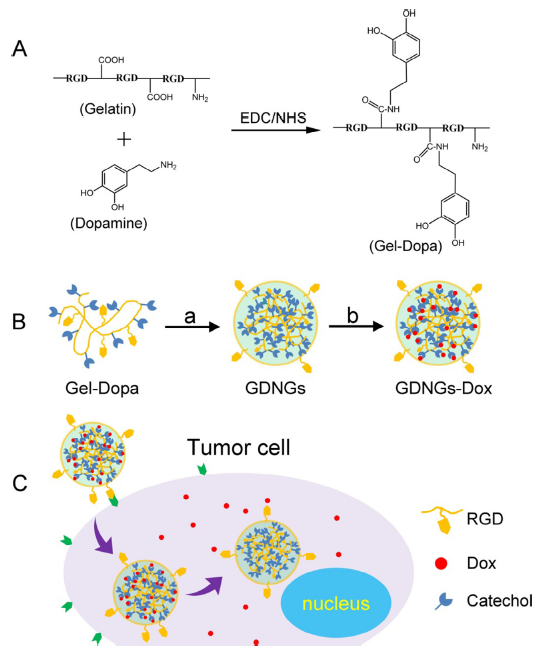
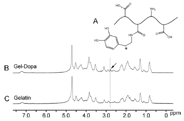
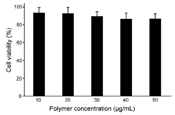


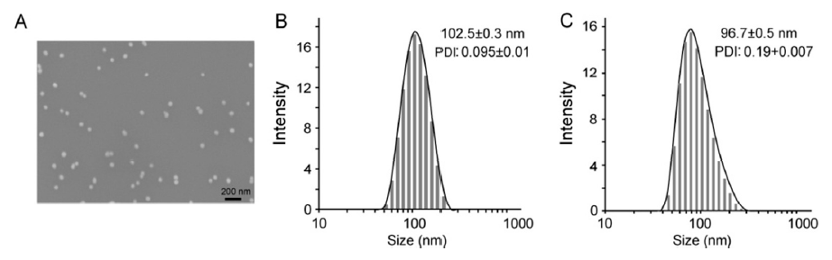
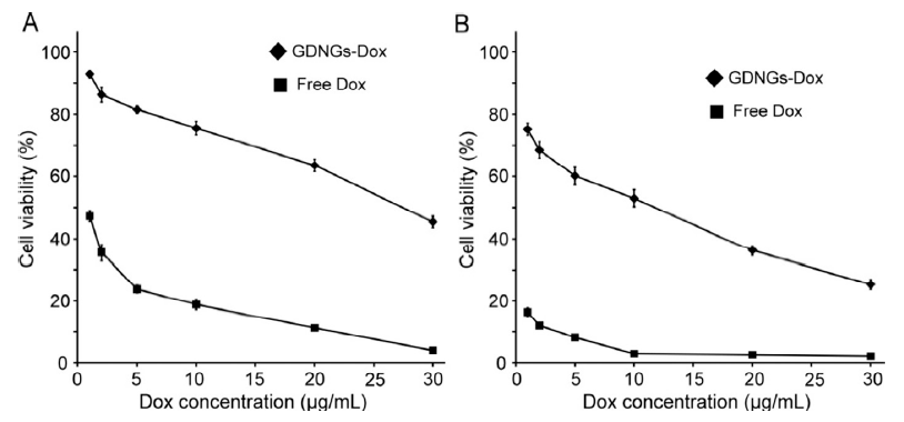