Search for articles:
Yanmei Zheng , Zhongtao Chen
Corresponding authors:
Received: 2019-06-24
Revised: 2019-08-26
Accepted: 2019-09-10
Online: 2020-03-15
Copyright: 2020 Editorial board of Journal of Materials Science & Technology Copyright reserved, Editorial board of Journal of Materials Science & Technology
More
Abstract
Graphitic carbon nitride (g-C3N4, CN) has attracted increasing interests in the field of photocatalysis due to its high visible-light-response. However, its photocatalytic activity is still lower for degradation of refractory contaminants such as Cr(VI) and Rhodamine B (RhB) etc. Herein, we report a facile method to synthesize a novel sulfur(S)-doped CN/reduced graphene oxide (rGO) porous nanosheet (S-CN/rGO PNs) via a supramolecular self-assembling followed by a solvothermal treatment. The as-prepared porous S-CN/rGO PNs are stable with high specific surface area ∼188.5 m2g-1 and exhibit a significantly enhanced photocatalytic activity of ∼17-fold and 15-fold higher than that of bulk CN for the degradation of RhB and Cr(VI) under visible light irradiation, respectively. Typically, 50 mL of 15 mg/mL RhB can be degraded within 20 min by 10 mg S-CN/rGO PNs. The mechanism can be explained by the synergistic effect of S doping and porous structure which can effectively reduce the band gap of CN and increase the specific surface area to promote the separation and transfer of photo-generated charge carriers. The results have provided a new way to significantly enhance the photocatalytic activity of g-C3N4 for degradation of refractory contaminants.
Keywords:
In recent years, the rapid development of industry and the extensive utilization of fossil fuels have caused environmental pollution, especially water pollution by organic conjugate compounds, inorganic poisonous and refractory heavy metals pollutants [[1], [2], [3]]. Photocatalysis has been considered as an intensely promising green technology to environmental remediation and clean energy production [[4], [5], [6]]. Graphitic carbon nitride (g-C3N4, CN) as a metal-free and high visible-light-response photocatalyst has drawn increasing interests for the potential application in hydrogen or oxygen evolution, environmental remediation, CO2 reduction, etc. due to its remarkable thermal chemical stability, facile synthesis route and moderate band gap (∼2.7 eV) [[7], [8], [9]]. However, the bulk CN has low specific surface area as well as rapid recombination rate of photo-generated electron-hole pairs and inefficient utilization of light energy. Quite a few strategies have been proposed to enhance its photocatalytic performance including thermal and liquid exfoliation [10,11], metal/non-metal doping [[12], [13], [14]], pore texture tailoring [[15], [16], [17]] and heterojunction [[18], [19], [20]] etc. Recently, two-dimensional ultrathin CN nanosheets with porous structure has attracted tremendous interests due to its tunable electronic structure, high surface area, and shorter charge migration length [21]. So far, a variety of porous CN has been synthesized through several typical methods, such as soft templating, hard templating, self-templating and template-free methods. Among them, self-templating strategies (e.g. supramolecular self-assembling) have appealed wide attention for the formation of hydrogen-bonded supramolecular assemblies between melamine precursors and triazine derivations. This has shown a key role in determining the different nanostructure morphology of porous CN materials [[22], [23], [24]].
In addition, recent reports have indicated that carbon materials loading is becoming one of the most promising strategy for improving the charge transfer and suppressing charge recombination. To date, various kind of carbon materials have been used to fabricate the CN/carbon complexes, such as CNTs [[25], [26], [27]], graphene [[28], [29], [30]], C60 [31], carbon fibers [32]. Graphene as a sp2-hybridized 2D carbon nanosheets has large specific surface area, high electrical conductivity and optical transmittance comparing with the 0D and 1D carbon materials [33]. Therefore, the combination of CN and graphene is considered to be one of the effective ways to improve the photocatalytic performance of CN. However, the bandgap of CN/graphene is still wide, resulting in a low visible light utilization.
Recent researches indicate that the non-metal doping of CN including S [[34], [35], [36]], P [37,38], B [39,40], C [41,42], etc. can narrow the bandgap of CN and enhance its light capture capability due to the chemical substituted doping [4]. Interestingly, the sulfur doping can promote the conductivity, mobility and separation of photo-generated electrons. It is believed that S atoms can achieve the replacement of N atoms [43]. Furthermore, it is well known that S atoms have been found to preferentially substitute N atoms with a large electronegativity, thus leading to the decreased of the band gap.
In addition, solvothermal treatment is an effective method to reduce GO and dope elements to CN owing to its high pressure and temperature. Furthermore, a porous structure can be constructed during solvothermal treatment because of the O2 pathway released by water vapor and the reduction of GO [44].
Herein, we report a facile method to synthesize a novel S-doped CN/rGO porous nanosheets (S-CN/rGO PNs) via a supramolecular self-assembling followed by a solvothermal treatment. The as-prepared S-CN/rGO PNs are stable with porous structure and exhibit an significant enhancement to the photocatalytic activity of CN for the degradation of RhB and Cr(VI) under visible light irradiation.
Typically, 5 g melamine was put into a 50 mL crucible and calcined at 550 °C for 2 h in muffle furnace. Then, the crucible was naturally cooled to room temperature to obtain yellow bulk CN.
2.5 g melamine powders and 2.55 g cyanuric acid were dissolved in 100 mL and 50 mL dimethyl sulfoxide (DMSO, sulfur source) under sonication, respectively. The obtained solutions were mixed together for 10 min to get white precipitates at room temperature. Subsequently, the mixture was centrifuged at 3000 rpm for 5 min to remove aggregates, and then washed with ethanol. The obtained white powder was dried at 60 °C overnight and calcined at 520 °C for 2 h with a heating rate of 5 °C min-1 in a muffle furnace to obtain curled light-brown CN PNs.
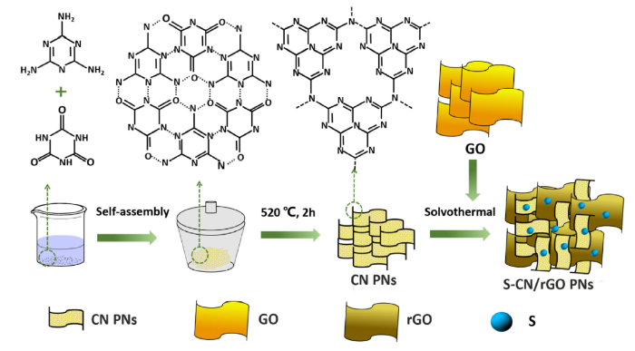
The S-CN/rGO PNs were prepared by solvothermal treatment of CN PNs and GO. Typically, 20 mg CN PNs were added into 50 mL DMSO. GO prepared by modified Hummers method [44] was added into the CN PNs solution with ultrasonic dispersion for 1 h. Then the mixed solution was transferred to a Teflon-lined stainless steel autoclave (100 mL) and heated at 180 °C for 12 h. At room temperature, the S-CN/rGO PNs were filtered to remove aggregates and washed with ethanol. The as-prepared composites were dried at 60 °C. The samples were labeled as S-CN/rGOx PNs (x = 2.5, 5 or 7.5, corresponding to the mass percentage of GO). For comparsion, S-CN PNs and CN/rGO NPs were also synthesized by the same solvothermal procedure as to get S-CN/rGO PNs except adding GO and DMSO, respectively. The preparation process and mechanism of melamine cyanuric acid complex (MCA) were formed by melamine and cyanuric acid via hydrogen bond self-assembly in DMSO. The nanoporous CN PNs were prepared by thermal polycondensation of MCA at 520 °C for 2 h in air. The porous S-doped S-CN/rGO PNs were prepared by solvothermal treatment of mixed GO, CN PNs and DMSO shown in Fig. 1.
Fig. 1. Schematically shown the preparation process and mechanism of S-CN/rGO PNs.
Scanning electron microscopy (SEM, Sirion) and Transmission electron microscopy (TEM, JEOL-2100 F) were used to study the morphologies and microstructures of samples. X-ray diffraction (XRD, D8-Discover) was used to identify the crystal structures and phase compositions of the samples by scanning at angles of 2θ = 10°-80°. The Brunauer-Emmett-Teller (BET, ASAP 2020) surface area was determined by nitrogen adsorption desorption isotherm. In addition, the pore size distribution of sample was calculated from the adsorption isotherm branch using the Barrett-Joyner-Halenda (BJH). Fourier transform infrared spectroscopy (FT-IR, Nicolet 5700 Fourier-IR spectrometer) was performed to study the chemical groups of samples. The chemical constitution was characterized by X-ray photoelectron microscope (XPS, PHI 500) and Raman spectra (Thermo Fish). Diffuse reflectance UV-vis spectra of photo catalysts were measured by a Shimadzu UV-2550 UV-vis spectrometer. The PL spectra were analyzed with a Shimadzu RF-5301 instrument at room temperature using a fluorescence spectrophotometer at excitation wavelength of 380 nm.
Photoeletrochemical measurements were performed using an electrochemical workstation (CHI660E, Shanghai, China) under a three-electrode system, i.e.: a S-CN/rGO PNs electrode, a Ag/AgCl electrode and a Pt foil as the working electrode, reference electrode and counter electrode, respectively. The working electrode was prepared by dispersing 1 mg phtocatalyst and 1 mL ethanol containing 40 u L Nafion with sonication. Then, 20 u L of the above solution was dropped onto ITO glass (20 × 20 × 2.3 mm3, 10 Ωsq-1) with an area of 100 mm2. The samples were dried at room temperature for 24 h. Before irradiation, the 0.1 moL-1 Na2SO4 solution was inlet with N2 to remove the electrolyte dissolved gas. The tests were conducted in an irradiation reaction vessel using a 300 W xenon lamp.
Photocatalytic activity was measured by using RhB, methyl orange (MO), methylene blue (MB) and Cr(VI) as the representative organic conjugate compound and inorganic heavy metal pollutant. The pollutants were dispersed in an aqueous solution under simulated solar light irradiation using a 300 W xenon lamp. Typically, 10 mg as-prepared photocatalyst was dispersed in 50 mL substrate aqueous solution in a cylindrical quartz vessel. Before irradiation, the suspension was magnetically stirred in dark for 0.5 h to ensure the adsorption-desorption equilibrium between substrate aqueous solution and photocatalyst. Then, 5 mL of the dispersion was taken at different time intervals during irradiation and then centrifuged to remove the photocatalyst. The concentration of RhB, MO, MB and Cr(VI) was examined by a Shimadzu UV-2550 UV-vis spectrometer. The 0.1 M HCl was used to adjust the pH value.
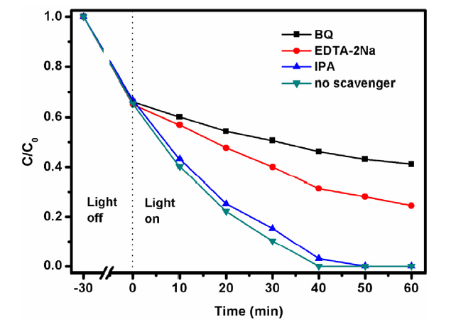
Activity species-trapping experiment was analyzed in RhB photodegration experiment by adding 1 mM ethylene diaminetetraacetic acid disodium salt (EDTA-2Na), 10 mM isopropanol (IPA), and 1 mM benzoquinone (BQ) as scavengers to quench holes (h+), hydroxyl radicals ($^{\cdot}$OH), and superoxide radicals ($^{\cdot}$O2-), respectively.
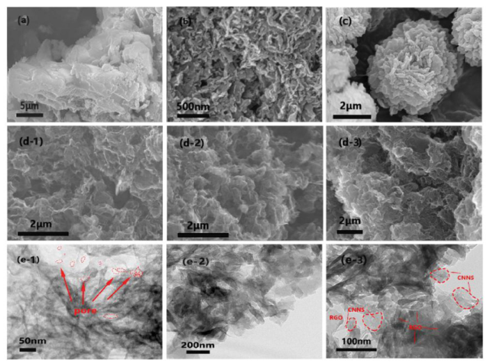
Fig. 2 shows the morphology of the as-prepared samples. In Fig. 2(a) and (b), CN shows a layered structure and CN PNs displays a curled nanosheet structure, respectively, caused by the precursor of the flower-like, layered spherical structure in Fig. 2(c). After calcination, the as-prepared CN PNs (Fig. 2(e-1)) exhibit porous structure, which is attributed to the release of a large amount of NH3 at high temperature. In the process of supramolecular self-assembling, the hydrogen-bonded is formed between melamine precursors and triazine derivatives [45]. The S-CN/rGOx PNs nanosheets structure can be confirmed by the TEM images of S-CN/rGO5 PNs in Fig. 2(e). The results imply that the structure is not affected by the solvothermal treatment, and the nanosheets structure can bring good electrical conductivity and short charge transferring length.
Fig. 2. SEM images of (a)-(c) CN, CN PNs and MCA with different magnification, and (d) S-CN/rGO2.5 PNs, S-CN/rGO5 PNs and S-CN/rGO7.5 PNs; TEM images of (e-1) CN PNs, and (e-2, e-3) S-CN/rGO5 PNs hybrid with different magnification.
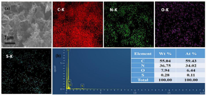
Fig. 3(a) shows the SEM image of S-CN/rGO5 PNs and the corresponding element mappings of C, N, O, S elements. It can be seen that all elements are homogeneously dispersed on the surface of S-CN/rGO PNs. It can be confirmed that S element is successfully doped into the CN PNs by the EDX spectrum in Fig. 3(b).
Fig. 3. EDX mapping of C (red), N (yellow), O (pink) and S (blue) elements (a), and EDX spectrum (b) of S-CN/rGO5 PNs.
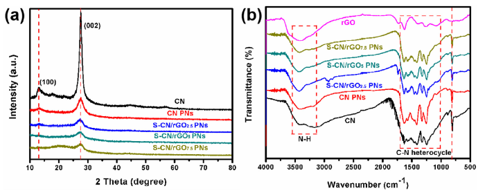
The crystal structure of the samples was characterized by XRD. As shown in Fig. 4(a), the XRD spectra show two typical diffraction peaks at 27.5° and 13.5°, respectively. The peak at 27.5° is a characteristic interlayers stacking reflection of conjugated aromatic systems, indexed for graphitic materials as the (002) peak, and the peaks at 13.5° is attributed to the (100) of the in-plane trigonal N linkage of tri-s-triazine motifs [46].
Fig. 4. XRD patterns (a), and FTIR spectra (b) of CN, CN PNs and S-CN/rGOx PNs.
In comparison with the CN, the intensity of (002) peak of the other samples is significantly decreased, demonstrating that the nanosheets structure have been successfully prepared. This is consistent with the results of SEM and TEM images. It is worth noted that the patterns of S-CN/rGO7.5 PNs exhibit a wide diffraction peak at 20°, represting a carbon diffraction peak because of complex containing of rGO. In addition, the peak at 13.5° becomes weaker in comparison with CN, which can be attributed to the sulfur doping into CN crystal lattice. The similar diffraction peaks also indicate that the solvothermal treatment and annealing have almost no effect on the crystallization of CN PNs and CN/rGOx PNs. The chemical structures of the samples were further confirmed by the FT-IR spectra as shown in Fig. 4(b). The characteristic peaks of CN at 810, 1200-1650 and 3000-3300 cm-1 are belong to the typical characteristic peaks of the s-triazine ring system, stretching modes of C—N heterocycles and the uncondensed amine group [47], respectively. The peaks of rGO can be further confirmed by Raman spectroscopy in Fig. S1. However, no peak is ascribed to the bond of sulfur, because the sulfur content in CN/rGOx PNs samples is low.
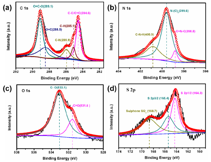
XPS measurement was performed to further investigate the chemical elemental compositions and valence states on the surface of S-CN/rGO5 PNs. As shown in Fig. 5(a), the C 1s XPS spectra can be deconvoluted into five bands of C—C/C=C peak at 284.6 eV, C—H peak at 285.1 eV, C—N peak at 285.9 eV, C=O peak at 286.6 eV, and O=C—O peak at 289.1 eV, respectively. These can be assigned to the graphite C—C bonds of the adventitious carbon atom, the sp2-hybridized carbon involved in the triazine rings (N—C=N), and the C—O bond formed by oxygen doping. Fig. 5(b) depicts the XPS peaks of N 1s, the peak at 400.9 eV is attributed to the C—N—H functional groups, hydrogen bond or charging effects.
Fig. 5. XPS spectra of S-CN/rGO5 PNs.
In addition, the binding energy of 399.6 eV is identified as N-(C)3 formed by tertiary N band to C. Finally, nitrogen species with binding energy of 398.8 eV is attributed to sp2-hybridized N (C=N—C) involved in triazine rings. The O 1s spectrum in S-CN/rGO5 PNs is showed in Fig. 5(c), the peak at 533.1 eV corresponds to the C—O bond. However, the peak at 531.6 eV is probably due to the surface absorbed oxygen when calcinated around the air. In addition, the S 2p spectrum is observed in Fig. 5(d), the peak located at 168 eV corresponds to the presence of sulfite species such as Ph-SO3- due to the oxygen from rGO and the sulfur from DMSO. The peak at 164.3 eV and 165.4 eV are ascribed to S 2p1/2 and S 2p3/2, respectively, which further confirmed that sulfur was successful doped into the S-CN/rGO PNs hybrid.
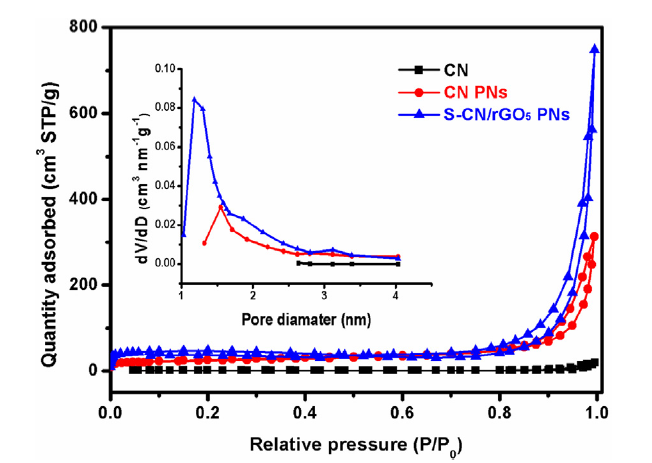
Combination with the EDX mapping, it can be confirmed that the S element has been doped into the S-CN/rGO PNs nanosheets hybrids. The textural properties was investigated by the nitrogen adsorption-desorption isotherm at 77 K as shown in Fig. 6. It can be seen that all samples can be categorized as type IV isotherms with H3 hysteresis loops. The adsorption isotherm shows a rapid increase when the relative pressure is close to unity, indicating the present of large mesoporous. Furthermore, the pore size is mainly less than 4 nm from the pore size distribution by BJH method (inset in Fig. 6), which is beneficial to the enhanced photocatalytic activity. The surface area of S-CN/rGO5 PNs is approximately 188.5 m2 g-1, which is about 39 times and 2 times higher than that of CN (4.885 m2 g-1) and CN PNs (92.3 m2 g-1). The result can further confirm that the increased surface area could be a significant factor for the potential enhanced photocatalytic activity.
Fig. 6. N2 adsorption-desorption isotherms and the corresponding pore size distribution (the inset) calculated by the BJH method for CN, CN PNs and S-CN/rGO5 PNs.
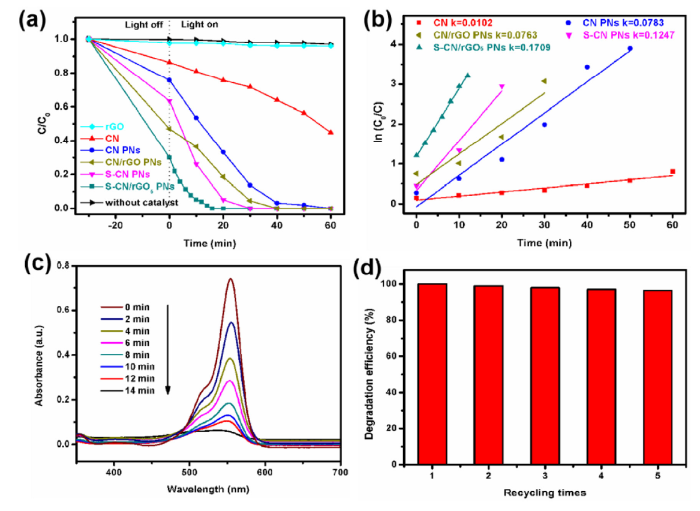
Fig. 7 represents the results of photocatalytic activity of the as-prepared samples. In Fig. 7(a), it can be clearly seen that the sample of S-CN/rGO5 PNs has the best photocatalytic activity, which degrades 100% of RhB within 20 min. In contrast, the CN, CN PNs, CN/rGO and S-CN/rGO PNs degrade 25%, 67%, 81% and 95%, respectively, indicating sulfur doping can be used to enhance the photocatalytic activity. With the increasing of rGO, the photocatalytic degradation for RhB gradually increase as shown in Fig. S2. However, further increasing the rGO in S-CN/rGO PNs, the degradation efficiency for RhB further decreases, which may be attributed to the light absorption competition between rGO and CN. Moreover, the rate constant of the S-CN/rGO5 PNs is about 17-fold of that CN under visible light irradiation, as shown in Fig. 7(b). Fig. 7(c) displays that the RhB solution is almost degraded by S-CN/rGO5 PNs within 14 min, which mainly attributed to the present of graphene and the large surface of the nanosheets structure. The cyclic stability of S-CN/rGO5 PNs was investigated by recycling the photocatalytic degradation tests. As shown in Fig. 7(d), no obviously decrease of stability activity is observed after 5 times of cycling tests, indicating that the sample of S-CN/rGO5 PNs possesses high stability.
Fig. 7. Photocatalytic degradation activity (a), and Photo degradation spectra (b) of RhB by as-prepared samples; (c) UV-vis spectra of RhB solution with S-CN/rGO5 PNs under sunlight; (d) Cyclic stability of S-CN/rGO5 PNs for photocatalytic RhB degradation (RhB concentration: 15 mg/L, photocatalyst dosage: 10 mg, volume:50 mL, irradiation time: 60 min, 5 cycles).
Cr(VI) was used as inorganic pollutants to evaluate the photocatalytic activity of the as-prepared catalysts under visible light irradiation. Fig. 8(a) shows the photocatalytic degradation rates of Cr(VI) aqueous solution over different samples. The introduction of rGO and S doping in S-CN/rGO5 PNs leads to the high specific surface area and more activity sites for the enhanced photocatalytic activity. Under the visible light irradiation, the S-CN/rGO5 PNs exhibit excellent photocatalytic degradation activity by removing 85.2% Cr(VI) within 100 min, while 73.9%, 51.5%, 36.9% and 14.2% Cr(VI) can be removed by S-CN PNs, CN/rGO PNs, CN PNs and CN, respectively. The results suggest that the doped of sulfur played a dominant role in photodegradation.
Fig. 8. (a) Photocatalytic degradation rates of Cr(VI) by as-prepared samples and without catalyst(C(K2Cr2O7) = 10 mg mL-1, pH = 3); (b) Photodegradation activity of Cr(VI) by S-CN/rGO5 PNs under different pH (C(K2Cr2O7) = 10 mg mL-1) and different Cr(VI) concentrations (c) (pH = 3); (d) Cyclic stability of S-CN/rGO5 PNs for photocatalytic degradation of Cr(VI) (C(K2Cr2O7) = 10 mg mL-1, pH = 3, 5 cycles).
The pH value is an important factor for photocatalysis performance. The pH value of Cr(VI) solution was varied from 2.0 to 6.0. Cr(VI) mainly exists in solution as Cr2O72-, CrO42- at low pH condition with the potential of E(Cr2O72-/Cr3+) ∼1.23 eV and E(CrO42-/Cr(OH)3) ∼ -0.13 eV. From Fig. 8(b), the rate of Cr(VI) degradation is enhanced by decreasing the pH value. The enhanced photocatalytic activity is attributed to the high E(Cr2O72-/Cr3+) at lower pH, which promotes the photo reduction.
Considering the excellent catalysis performance in acid condition, the pH = 3 was chosen for further study. As shown in Fig. 8(c), the effect of Cr(Ⅵ) concentration on photo reduction performance of S-CN/rGO5 PNs hybrid was also investigated. It can be observed that 100% Cr(VI) aqueous solution can be removed in the concentration of 5 mg L-1 within 40 min, while 85.2%, 57.1%, 52.4% and37.9% for 10 mg L-1, 20 mg L-1, 40 mg L-1 and 60 mg L-1, respectively. The stability and reliability of the catalyst are critical to the practical application. As shown in Fig. 8(d), it can be observed that the S-CN/rGO5 PNs still retain a removal rate of 59.1% after five cycles, showing an excellent photocatalytic cycling ability. Fig. S3 shows the XPS spectra of S-CN/rGO5 PNs after five cycles of Cr(VI) photoreduction. The binding energy peaks at 577.8 eV and 587.4 eV are attributed to the Cr(Ⅲ) 2p3/2 and Cr(Ⅲ) 2p1/2, respectively, which confirms the reduction of Cr(Ⅵ) to Cr(Ⅲ) on the S-CN/rGO5 PNs surface.
To further test the photocatalytic ability of the as-obtained catalyst, the degradation of MO and MB were studied shown in Figs. S5 and S6. Above all, the prepared S-CN/rGO PNs sample exhibit highly efficient photocatalytic degradation of refractory contaminants due to the introduction of rGO and S.
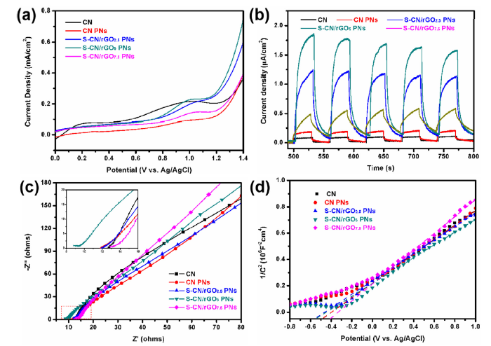
Photoelectrochemical measurements are used to investigate the excitation and transfer of photogenerated electro-hole pairs in photocatalysis to understand the possible photocatalytic mechanism. Fig. 9(a) shows the linear sweep voltammograms (LSV) curves of CN, CN PNs and CN/rGOx PNs samples. Due to the synergistic effect of S-doping and rGO, the oxygen evolution potential moves negatively, resulting in the rapid separation of electron-hole pairs and more oxidation catalysis centers formed by S and rGO. This is beneficial for absorbing more oxygen. The transient photocurrent responses recorded for several on-off cycles under irradiation of simulated sunlight of CN, CN PNs and CN/rGOx PNs are shown in Fig. 9(b). The CN/rGOx PNs shows the highest photocurrent intensity due to its large specific surface area and high conductivity to enhance the separation efficiency of electron-hole pairs. The S-CN/rGO2.5 PNs, S-CN/rGO5 PNs and S-CN/rGO7.5 PNs are about 15.4, 23.1 and 6.0 times higher than that of CN and 6.9, 10.3 and 2.7 times higher than that of CN PNs, respectively. The result indicates that doping too little or too much rGO will cause decreased current density, which can be attributed to the negative shading effect of rGO on CN PNs [48]. The shading effect implies that overloading of rGO will affect the light transmission of the samples and the active sites of CN PNs. The Nyquist plots of EIS spectra of these samples are displayed in Fig. 9(c). The radius of the impedance arc is mainly in direct proportion to the charge transfer resistance. The S-CN/rGO5 PNs possesses the smallest semicircle and internal resistance as shown in the inset of Fig. 9(c). This indicates that the charge transfer resistance is obviously decreased. The Mott-Schottky method is employed to measure the semiconductor property of these samples as shown in Fig. 9(d). We can infer that all samples show n-type characteristics because the slopes of liner region are positive. The small slope value of tangent line of S-CN/rGO5 PNs can be attributed to the reason that the conductivity of the composites is greatly affected by the doping levels, which is consistent with LSV and transient photocurrent responses.
Fig. 9. (a) LSV curves, (b) Transient photocurrent responses, (c) EIS Nyquist plots, and (d) Mott-Schottky plots of CN, CN PNs and CN/rGOx PNs obtained in 0.1 M Na2SO4 electrolyte under irradiation of simulated sunlight.
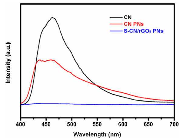
The photoluminescence (PL) spectra at room temperature are deemed as an efficient way to prove the efficiency of electron-hole pair trapping, migration and transfer. The PL spectra of CN, CN PNs and S-CN/rGO5 PNs are shown in Fig. 10. The major emission bands of CN and CN PNs are 460 nm and 450 nm, respectively. The slightly red‐shift ∼10 nm of the major emission bands is attributed to the well-known delamination induced quantum confinement effect [49]. The PL intensity of S-CN/rGO5 PNs decreases fast that of CN and CN PNs. This can be attributed to the formation of hydrogen bonds facilitates efficient energy transfer between molecules and delocalization, which enhance the ability of the catalyst to capture visible light [13].
Fig. 10. PL spectra of CN, CN PNs and S-CN/rGO5 PNs excitated at 370 nm.
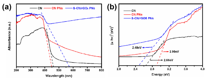
The UV-vis absorption spectra, see Fig. 11, are used to investigate the electronic band structure of the samples. In Fig. 11(a), CN exhibits typical semiconductor absorption with band edge position at about 460 nm as a result of the charge transfer response from the N 2p orbitals to the C 2p orbitals. In addition, the absorbance spectrum of S-CN/rGO5 PNs shows an increased light absorption at about 570 nm, which is related to the sulfur doping and the high conductivity of rGO. However, CN PNs show a slightly blue-shift in comparison with CN, attributed to the quantum confinement effect [13]. Based on these results, the photogenerated electron-hole pairs inside the CN PNs can be rapidly transferred to the surface in the interlayer direction. The photogenerated electron-hole pairs on the surface of the CN PNs can be rapidly moved to the reaction sites along the inner layer. Obviously, the inherent characteristics of CN PNs will help to achieve the maximum improvement of photocatalytic performance. The optical band gap (Eg) of all samples were determined from the (ahν)2 versus photon-energy plots shown in Fig. 11(b). It reveals a band gap of about 2.66 eV, 2.90 eV, and 2.48 eV of CN, CN PNs and S-CN/rGO5 PNs, respectively.
Fig. 11. UV-vis absorption spectra (a) and UV-vis diffuse reflectance spectroscopy (b) of CN, CN PNs and S-CN/rGO5 PNs.
The trapping experiments were used to identify the active species during the photocatalytic process. As scavengers, EDTA-2Na, IPA and BQ were added to quench the h+, ·OH and ·O2-, respectively. The result show that, the biggest suppression occured when BQ was added shown in Fig. 12, implying that $^{\cdot}$O2- is the main active species in the photocatalytic reaction. Meanwhile, once EDTA-2Na was added, the degradation rate of RhB observed a decrease, indicating that h+ also played a dominant role in RhB photodegradation. However, slightly decrease of RhB degradation rate was induced when IPA was added, suggesting that $^{\cdot}$OH was not the crucial species to the RhB photodegradation.
Fig. 12. Photodegradation of RhB in active species trapping experiment with S-CN/rGO PNs as photocatalyst (RhB concentration: 20 mg/L, photocatalyst dosage: 10 mg, volume: 50 mL).
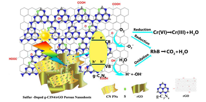
Base on the above results, a possible mechanism for the photocatalytic degradation of RhB and Cr(VI) by S-CN/rGO5 PNs photocatalyst is proposed in Scheme 1. As shown in Scheme 1, under visible light irradiation, the stimulated CN can cause the electron (e-) transition from the valence band (VB) to the conduction band (CB), leaving a hole (h+) on the VB (Eqs. (1)). Meanwhile, the photo generated electrons on the CB can be further transferred to the surface and easily reacted with surface O2 to produce superoxide ($^{\cdot}$O2-) (Eqs. (2)). The left holes in VB can react with the absorbed water to produce surface hydroxyl radicals ($^{\cdot}$HO-) (Eqs. (3)). Due to the excellent electrical conductivity of rGO, the generated carriers can be rapidly accepted and separated, which retards the recombination of electrons and holes. The strong oxidizing properties of $^{\cdot}$O2- can effectively degrade RhB (Eqs. (4)) and the $^{\cdot}$HO- can directly oxidize RhB (Eqs. (5)). Furthermore, the $^{\cdot}$O2- can reduce Cr (VI) to Cr (III) by the photogenerated electrons (Eqs. (6), (7)). The mesoporous of the nanosheets can act as a charge channel to shorten the transmission distance of the electrons from internal phase to surface. Benefiting from the stacking distance of the interlayer of CN and rGO as well as the sulfur impurities, S-CN/rGO5 PNs hybrid possess an excellent photocatalytic performance.
g-C3N4+hν→h +e- (1)
O2 +e-→$^{\cdot}$O2- (2)
h+ + H2O→$^{\cdot}$OH+H+ (3)
RhB+$^{\cdot}$O2- → CO2 +H2O (4)
RhB+$^{\cdot}$OH→CO2 +H2O (5)
Cr2O72-+14H+ + 6e- → 2Cr3+ + 7H2O (6)
CrO42- + 8H+ + 3e- → Cr3+ + 4H2O (7)
Scheme 1. Schematic showing the mechanism of RhB degradation by S-CN/rGO5 PNs hybrid.
We have developed a facile method to synthesize a novel S-CN/rGO PNs via a supramolecular self-assembling followed by a solvothermal treatment. The as-prepared S-CN/rGO PNs are stable with porous structure and exhibit a significantly enhanced photocatalytic activity for the degradation of RhB and Cr(VI) under visible light irradiation, respectively. The mechanism can be explained by the synergistic effect of S doping and porous structure which can effectively reduce the band gap of CN and increase the specific surface area to promote the separation and transfer of photo-generated charge carriers. The results have provided a new way to significantly enhance the photocatalytic activity for degradation of refractory contaminants.
The authors would like to thank the Science and Technology Major Project of Shanxi Province (Grant Numbers MC2016-06), National Natural Science Foundation of China (21173041) and the Opening Project of Jiangsu Key Laboratory of Advanced Metallic Materials, China.
Supplementary material related to this article can be found, in the online version, at doi:https://doi.org/10.1016/j.jmst.2019.09.018.

 WeChat
/
| 〈 |
|
〉 |
