Search for articles:
Peng Chen , Tianxing Chen
Corresponding authors:
Received: 2019-02-11
Revised: 2019-03-16
Accepted: 2019-05-21
Online: 2019-10-05
Copyright: 2019 Editorial board of Journal of Materials Science & Technology Copyright reserved, Editorial board of Journal of Materials Science & Technology
More
Abstract
In order to develop a facile and precisely controlled approach to synthesize hierarchical mesoporous materials with tailored property, in this work, a novel study was carried out to fabricate montmorillonite-chitosan hollow and hierarchical mesoporous spheres (MMTNS@CS-HMPHS) based on single-template layer-by-layer (LbL) assembly. Scanning electron microscopy (SEM), transmission electron microscopy (TEM), specific surface area analysis and X-ray photoelectron spectroscopy (XPS) analyses were carried out to characterize the morphology and surface properties of MMTNS@CS-HMPHS. Benefitting from the unique lamellar structure of MMTNS, mesoporous channels are formed on the shell of MMTNS@CS hollow spheres, resulting in high surface area. Moreover, the surface functionalization and pore size of MMTNS@CS-HMPHS can be easily tuned, due to the tailored property through LbL assembly method. Besides the unique microstructure, MMTNS@CS-HMPHS also possesses the active sites generated from both MMT and chitosan, which greatly promotes its performance in fields of adsorption, drug delivery and catalyst supports, etc.
Keywords:
As a kind of porous material with unique morphology, hollow-structured materials have shown many promising application prospects in various fields, such as adsorption and catalytic, storage, targeted drug delivery, etc. [1,2], primarily attributed to its merits of low density, large void space, and large specific surface area [3,4]. Additionally, in order to further promote the exchange of substances and reduce transport limitations, considerable efforts have been devoted to fabricate hollow sphere with a hierarchical mesoporous structure [5,6].
According to the template synthesis concept for hierarchical mesoporous materials, one of the most common method is to construct hierarchical mesoporous system by using two templates of different molecular weight (copolymer and surfactant pairs) via self-assembly [7]. Niu et al. [5] successfully synthesized hierarchical mesoporous silica spheres by utilizing an amphiphilic block copolymer (polystyrene-b-poly (acrylic acid)) and cetyl trimethyl ammonium bromide as co-templates. Rana et al. [8] prepared hierarchical mesoporous bio-polymer/silica composite materials using a dual-template of the cationic N, N, N-trimethyl chitosan and the anionic sodium dodecyl sulfate. Although the thickness of the shells and the pore size in the core could be easily tuned by changing the template molecules, the assembly process is still difficult to control, for the template molecules tend to form either mixed micelles or separated macroscopic phases during the preparation process [9]. Therefore, developing a facile and precisely controlled approach to synthesize hierarchical mesoporous materials with tailored properties is necessary.
LbL assembly is a simple, highly versatile, precisely controlled approach that has been widely used to prepare nanostructured materials with tailored properties [10,11]. The most obvious superiority of LBL assembly is that the properties of products, such as thickness, composition, and function, can be readily tuned by simply varying the number of layers deposited, the type of species adsorbed, and the conditions employed during the LbL assembly process [12]. Thus, in this work, montmorillonite nanosheets and chitosan hollow sphere with hierarchical mesoporous (MMTNS@CS-HMPHS) was directly synthesized through single-template LbL assembly.
Montmorillonite (MMT) is one of the most abundant natural clay minerals with 2D sheet-like morphology. Benefitting from its distinctive swelling and cation exchange properties [13], MMT is widely used as adsorbents, ion-exchange reagents, catalyst supports, and additives for polymer nanocomposites [14,15]. Nevertheless, the physicochemical properties of MMT can be further improved through forming a unique hierarchical mesoporous structure.
In this work, positively charged chitosan and negatively charged MMTNS were alternatively deposited on the surface of polystyrene (PS) spheres through electrostatic interaction via LbL assembly, followed by removing the PS spheres. The obtained product was characterized by transmission electron microscopy (TEM) and scanning electron microscopy (SEM), which demonstrated the successful preparation of MMTNS@CS-HMPHS.
MMT was obtained from Ningcheng Tianyu Bentonite Technology Co., Ltd. (Inner Mongolia, China). Negatively charged PS spheres with a diameter of 800 nm were purchased from Shangwei Biological Technology Co., Ltd. (Jiangsu, China). All other regents used in this experiment were purchased from Sinopharm Chemical Reagent Co. Ltd. (Shanghai, China). The water used in this work was produced by a Millipore Milli-Q Direct 8/16 water purification system with 18.2 MΩ.
The preparation of MMTNS was got according to an ultrasonic process rather similar to that introduced in our previous work [16,17]. MMT suspension was treated by a Cole Parmer ultrasonic processor (750 W and 20 kHz) with 60% amplitude for 4 min. Finally, the colloidal suspension was centrifuged at 13,000 rpm for 4 min to remove the unexfoliated product, and the homogeneous supernatant was MMTNS suspension.
The synthesis route of MMTNS@CS-HMPHS is shown in Fig. 1, firstly, a total of 50 mg PS spheres was added into 50 mL of chitosan solution (0.5%) and stirred for 5 min, then rinsed with deionized water to remove excess chitosan. Subsequently, the chitosan coated PS spheres were soaked into MMTNS suspension (0.5%) for 5 min, followed by rinsing and freeze-drying. Finally, MMTNS@CS-HMPHS was obtained through calcination in N2 at 500 °C for 2 h to remove the PS core.
Fig. 1. (a) Schematic illustration of the route for preparing MMTNS@CS-HMPHS and (b) unique microstructure and potential applications of MMTNS@CS-HMPHS.
The thickness of MMTNS was obtained under ambient conditions using an atomic force microscope (AFM; MultiMode 8, Bruker). The morphology of the samples was observed using a SEM (Sigma300, Carl Zeiss) at an accelerating voltage of 2 kV. TEM test was carried out on a JEM-2100F microscope operating at 200 kV. X-ray photoelectron spectroscopy (XPS) measurements were taken using an ESCALAB 250Xi spectrometer. Nitrogen gas adsorption-desorption isotherms were obtained at 77 K using a TriStarⅡ3020 surface area analyzer.
Fig. 2(a) shows a typical AFM morphology image of the prepared MMTNS. Although several sheets are white, the color of most sheets is closer in brown, which indicates the MMT after ultrasonic treatment has a uniform thickness distribution. And the cross-section analysis along the red line in the AFM image of some brown sheets is shown in Fig. 2(b). The thickness of these sheets is about 1 nm, which is in accordance with the theoretical thickness of monolayer MMT [18], indicating MMTNS is well prepared through ultrasonic treatment.
Fig. 2. (a) Typical AFM morphology image (4 μm × 4 μm, 512 pixels) of the prepared MMTNS and (b) topographic profile along the red line in the corresponding image.
In order to construct MMTNS@CS-HMPHS, MMTNS and chitosan were deposited only single layer on PS sphere through LbL assembly, and the deposition process was monitored by SEM. For the original PS spheres (Fig. 3(a)), they have a regular morphology and uniform particle size distribution of about 800 nm. It is obvious that MMTNS successfully deposit on the PS surface via LbL assembly with chitosan, forming a core-shell structure, as shown in Fig. 3(b) and (e). Although the formed shell exists some defects that are gaps between MMTNS (Fig. 3(f)), and it is these defects that form mesoporous channels on the shell of MMTNS@CS hollow sphere in following calcination process. Furthermore, the morphology of PS@chitosan and PS@chitosan@MMTNS was investigated by TEM measurement. The results are shown in Fig. 3(c) and (d), chitosan and MMTNS successfully deposit on the surface of PS sphere through the strong contrast between the bright edge and the relatively dark center. The thickness of chitosan and chitosan@MMTNS shell is roughly estimated to be around 1 nm and 3 nm from the enlarged TEM image of PS@chitosan (Fig. 3(g)) and PS@chitosan@MMTNS (Fig. 3(h)), respectively.
Fig. 3. (a) SEM images of original PS spheres, (b, e, f) SEM images of PS spheres after LbL assembly with different magnification and (c, g, d, h) TEM images of PS@chitosan and PS@chitosan@MMTNS with different magnification, respectively.
To achieve a hollow architecture, calcination was utilized to remove the PS sphere template. The surface chemical composition and chemical states of MMTNS@CS-HMPHS were analyzed by XPS. From the atomic ratio in Table 1, both the elements of C, N that from chitosan and the elements of Al, Si, O, Mg that from MMT can be clearly identified from MMTNS@CS-HMPHS, which reveals the successfully composite of MMTNS and chitosan. The elementary composition of MMTNS@CS-HMPHS also can be obviously verified by the results of EDS elemental mapping (Fig. 4). It can be seen that the elements of Al, Si, O and C are distinctly detected and the intensity of Al, Si, Mg and O that from MMTNS is quite higher than the elements of C that from chitosan, which also reveals the successfully wrap of MMTNS to chitosan. Binding energies found at 284.59 eV and 286.42 eV for C 1s in MMTNS@CS-HMPHS (Fig. 5(a)) were attributed to C—C and C—O bonds of chitosan [19]. For O 1s spectrum (Fig. 5(b)), the peaks at 531.64, 532.19, and 533.64 eV can be assigned to O—C bonds from chitosan, the lattice oxygen (O2-) from MMT [20], and hydroxyl groups (OH-) from chitosan [21], respectively. This result reveals that MMTNS@CS-HMPHS possesses the surface properties of both MMT and chitosan, greatly promoting the application potential of MMTNS@CS-HMPH.
Table 1 Atomic fraction (%) of MMTNS@CS-HMPHS.
| O | Si | Mg | C | Al | N | Others |
|---|---|---|---|---|---|---|
| 46.15 | 19.23 | 2.3 | 20.74 | 7.03 | 1.39 | 3.16 |
Fig. 4. EDS elemental mapping of MMTNS@CS-HMPHS.
Fig. 5. XPS high-resolution scans for (a) C 1s and (b) O 1s of MMTNS@CS-HMPHS.
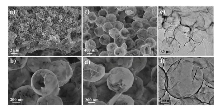
SEM images of the final MMTNS@CS-HMPHS with different magnification are shown in Fig. 6. As expected, the final MMTNS@CS-HMPHS still keeps regular morphology, and almost no collapse is observed after calcination. This is probably attributed to the tightly wrapped of MMTNS and chitosan, which hampers the deformation and maintains the spherical structure [22]. From the magnified SEM images of the broken MMTNS@CS-HMPHS (Fig. 6(c)), it can be seen that the PS sphere template is absolutely removed, and MMTNS@CS-HMPHS indeed has a hollow architecture. The morphology and microstructure of MMTNS@CS-HMPHS also can be clearly demonstrated by TEM images. As shown in Fig. 6(e) and (f), the strong color contrast between the dark edge and the relatively bright center is a direct evidence for the formed hollow structure. Furthermore, as shown in Fig. 6(d), there are some irregular mesoporous channels with a diameter of about 20 nm on the shell of MMTNS@CS hollow sphere. These results indicate that MMTNS@CS-HMPHS is successfully synthesized based on single-template LbL assembly.
Fig. 6. SEM and TEM images of MMTNS@CS-HMPHS with different magnification.
The surface area and pore structure of MMTNS@CS-HMPHS were investigated by N2 adsorption-desorption isotherms. The N2 adsorption-desorption isotherm curve of MMHTNS-HS (Fig. 7(a)) exhibits a type IV adsorption branch with a H3 hysteresis loop, which is the characteristic of mesoporous structure. The Brunauer-Emmett-Teller specific surface area and total pore volume of MMTNS@CS-HMPHS are calculated to be 81.32 m2 g-1 and 0.1944 cm3 g-1, respectively. Benefitting from its unique hierarchical mesoporous structure, the specific surface area of MMTNS@CS-HMPHS is quite higher than that of MMTNS (38.02 m2 g-1) [23]. Furthermore, there is a sharp peak at 4 nm in the pore-size distribution of MMTNS (Fig. 7(b)), which can be attributed to the narrow gaps of MMTNS [24].
Fig. 7. (a) Nitrogen sorption isotherm and (b) pore size distribution of MMTNS@CS-HMPHS.
The synthesized MMTNS@CS-HMPHS indeed has a hierarchical mesoporous structure (Fig. 1(b)), which greatly increases the effective surface area and promotes the exchange of substances between the internal and external surfaces. Therefore, this unique structure makes MMTNS@CS-HMPHS to be an ideal candidate for various applications including adsorption, catalyst supports, and drug delivery. In addition, the internal of MMTNS@CS-HMPHS is decorated by chitosan that possesses high contents of amino functional groups [25], which may further promote the performance of MMTNS@CS-HMPHS.
Interestingly, many hollow spheres are also synthesized through hard-templating and LbL assembly, but no mesoporous channels is found on their shell [26,27]. However, in this work, the synthesized MMTNS@CS hollow spheres indeed have hierarchical mesoporous structure. The primary difference of synthetic methods is that our raw material has a lamellar structure, while their raw materials are just nanoparticles. Based on this difference and above observations, a possible mechanism was proposed to explain the formation of mesoporous channels (Fig. 8). During the deposition process, MMTNS inevitably overlaps on the PS surface, due primarily to its unique lamellar structure. Then in the next step of rinse, the overlapped MMTNS drops from PS surface, owing to the weaker electrostatic attraction between MMTNS and chitosan. Finally, some defects are formed on the shell of MMTNS@CS hollow sphere (Fig. 8(d)). However, it is these defects that form mesoporous channels on the shell of MMTNS@CS hollow sphere after calcination. Furthermore, according to the proposed mechanism, the pore size of mesoporous channels can be easily tuned by changing the lateral size of MMTNS.
Fig. 8. Schematic illustration for formation of mesoporous channels on the shell of MMTNS@CS hollow sphere.
MMTNS@CS-HMPHS was successfully fabricated based on single-template LbL assembly. The surface functionalization and pore size of MMTNS@CS-HMPHS can be easily tuned by adding functionalized materials and changing the lateral size of MMTNS benefiting from the tailored property of LbL assembly. Furthermore, prepared MMTNS@CS-HMPHS has a distinctive hierarchical mesoporous structure and active sites of both MMT and chitosan, which offers greatly potential applications in fields of adsorption, drug delivery and catalyst supports, etc.
This work was supported financially by the National Natural Science Foundation of China (Nos. 51874220 and 51674183), the Natural Science Foundation of Hubei Province of China (No. 2018CFB468) and the Excellent Dissertation Cultivation Funds of Wuhan University of Technology (No. 2018-YS-050).

 WeChat
/
| 〈 |
|
〉 |


