Cytotoxic Effect on Osteosarcoma MG-63 Cells by Degradation of Magnesium
Mei Li1, a , Ling Ren2, a , LiHua Li1 , Peng He1 , GuoBo Lan1 , Yu Zhang1, *  , Ke Yang
2, **
, Ke Yang 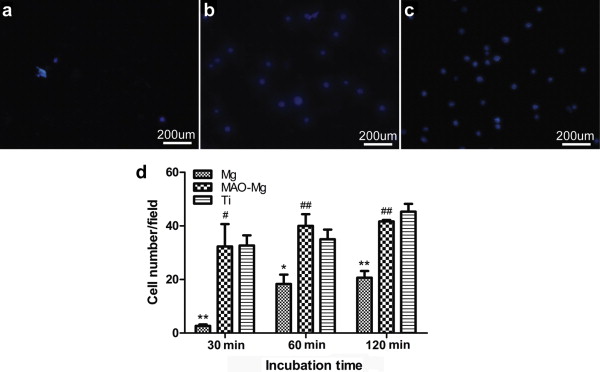
Cytotoxic Effect on Osteosarcoma MG-63 Cells by Degradation of Magnesium |
|
Mei Li
, Ke Yang |
| Fig. 5. Fluorescence micrographs of DAPI stained MG-63 cells after 30, 60 and 120 min incubations on different samples: a pure Mg; b MAO coated pure Mg; c pure Ti. The upper pictures showing images of cells attached on surfaces after 60 min of incubation. One symbol, p 0.05; two symbols, p 0.01. *Compared to Ti group at the same day, #: compared to Mg group with at the same day. |
| |