Cytotoxic Effect on Osteosarcoma MG-63 Cells by Degradation of Magnesium
Mei Li1, a , Ling Ren2, a , LiHua Li1 , Peng He1 , GuoBo Lan1 , Yu Zhang1, *  , Ke Yang
2, **
, Ke Yang 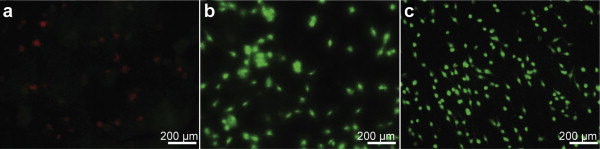
Cytotoxic Effect on Osteosarcoma MG-63 Cells by Degradation of Magnesium |
|
Mei Li
, Ke Yang |
| Fig. 2. Fluorescence micrographs of livedead dye-stained MG-63 cells after 24 h incubation on the sample surfaces: a pure Mg; b MAO coated pure Mg; c pure Ti. The living and dead cells are visualized in green and red by fluorescence microscopy, respectively. |
| |