Microstructural Evolution of a Ni-base Alloy DZ468 Joint Bonded with a New Co-base Filler
Jing Yanhong, Zheng Zhi, Liu Enze*  , Guo Yi
, Guo Yi 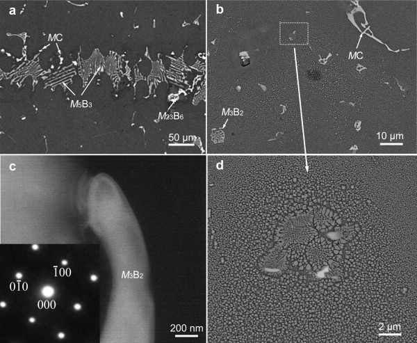
Microstructural Evolution of a Ni-base Alloy DZ468 Joint Bonded with a New Co-base Filler |
|
Jing Yanhong, Zheng Zhi, Liu Enze
, Guo Yi |
| Fig. 4. Images from the joint bonded at 1553 K for 5 min: a SEM micrograph of the bonding zone, b SEM micrograph of the diffusion affected zone, c morphology of Cr-rich boride M 3 B 2 in the liquid pool, the inset is the SAED pattern of the phase showing [001] zone axis, d micrograph of the liquid pool in the bonding zone. |
| |