

Rapidly Solidified Microstructure in Laser Alloyed Ni-Al Layer by TEM, STEM z-contrast and HRTEM Techniques
[Yue Yang1  , Jiandong Hu
2 ]
, Jiandong Hu|
|


In the present study, laser alloying of electroless Ni-P coating on aluminum substrate was conducted using Nd:YAG pulsed laser under the condition of 5.36 × 10⁹ W/m2 in power density and 3.0 mm/s in scanning speed. The rapidly solidified microstructure in the alloyed layer was studied. The results showed that the alloying element distributed in the alloyed layer is inhomogeneous. The dendrite containing relatively high Ni was identified as Al3Ni phase and the areas between the dendrites are rich in Al content. Featureless with cell structure in Al-rich areas was firstly displayed by z-contrast image. Amorphous structure was revealed to exist in Al-rich areas.
Laser alloying has been proven to be an effective method for the enhancement of hardness and the improvement of wear resistance of aluminum alloys, since the bonding of alloyed layer with substrate materials is a metallurgical process[1], [2] and [3]. In laser surface alloying, the most important factors are distribution of the alloying elements and the development of microstructure in the alloyed layer because they are closely related to the properties of the alloyed layer[4], [5] and [6]. Although some studies concerning laser alloying of Ni-P coating with aluminum substrate were reported, i.e. Ni-P amorphous phases formed in the laser processed Al with electroless deposited Ni-P coating and the effects of which on corrosion resistance were studied in our previous studies[7], [8] and [9]. New results can also be expected because of more advanced analysis techniques such as HRTEM (high resolution transmission electron microscopy) are used in the present study. Therefore, the purpose of this work is to study rapidly solidified microstructure in details by means of TEM (transmission electron microscopy), STEM (scanning transmission electron microscopy), z-contrast and HRTEM techniques. In the present work, a detailed microstructure characterization by TEM was carried out on the Ni-Al laser alloyed layer and an identification of phases in the coating was accomplished to reveal the details in the element and amorphous phase distributions.
The material used for laser alloying was commercial-purity aluminum with dimensions of 10 mm × 30 mm × 3 mm. Before laser alloying processing, Ni-P coating was chemically deposited on aluminum substrate. Prior to the deposition, the surfaces of Al plates were mechanically polished and then activated using HNO3 solution. Finally, the galvanizing treatment was conducted to the specimens for about 1 min. The bath solution containing 15 g/l NiSO4; 14 g/l Na2H2PO2; 13 g/l NaC2H3O2 and 30 g/l boric acid was employed to coat the samples, operated at 70 ° C and the pH value was kept as constant at 5. The deposition thickness of the coating on the samples was about 25 μ m. The composition of the coating determined by EDS (Model JSM-5310, Japan) was 6.74 at.% P and Ni balance.
Laser treatment was conducted on a Nd-YAG pulsed laser with the following parameters: power density of 5.36 × 10⁹ W/m2; scanning speed of 3.0 mm/s; duration time of 4 ms; spot size of 480 μ m. During the test, the sample surface was protected by a protective argon gas. After the laser treatment, selected specimens were sectioned and polished.
Microstructures and elements distribution analysis were examined by SEM (JSM-5600, Japan), TEM (model JSM-2100F, Japan) and EDS. During the procedure for preparing cross-sectional TEM specimens, the rectangular areas of treated layer are cut from bulk material parallel to the beam moving direction. Two sections of the treated layer are coated with epoxy, stacked face to face and cured under pressure to obtain a strong bond with minimum glue thickness (< 1 μ m). A cylinder is cut from the stack, inserted into a brass tube which is full of Gatan glue for final sectioning and eventually dimpling prior to ion milling. Microanalysis was carried out by HRTEM, with EDS and STEM z-contrast system attached.
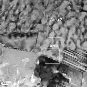 | Fig. 1 Laser alloyed layer revealed by TEM in cross section. |
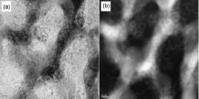
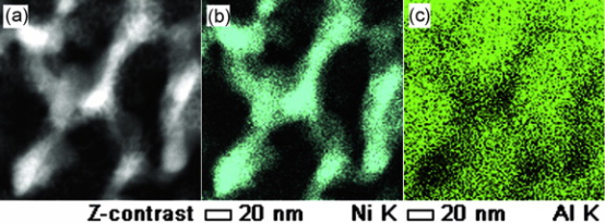 | Fig. 2 Laser alloyed dendrite structure revealed by z-contrast with HAADF (a), Ni distribution with EDS (b) and Al distribution with EDS (c). |
Fig. 3, BF and HAADF images in high magnification taken from the same area as that for Fig. 2, shows the details for the solidified structure. More fringes exist in dendrite as shown in Fig. 3(a), which may be resulted from coherent diffraction of Ni atoms. It is noted that Al-rich region includes a great deal of cell structure. Cell boundaries show high intensity in z-contrast image (Fig. 3(b)), indicating the segregation of high-Z atom such as Ni or other impurities. It is interesting to note that Al-rich area is in featureless.
 | Fig. 3 Laser alloyed dendrite structure reveled by (a) BF and (b) HAADF in high magnification. |
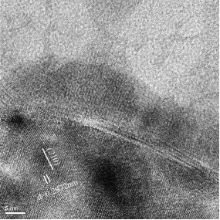
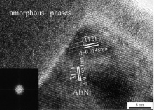
Fig. 4, an HRTEM micrograph taken from the area marked with square 2 in Fig. 1, shows crystal lattice image and featureless morphology at the same time, while the former corresponds to Ni-rich dendrite and the latter corresponds to featureless structure in Al-rich areas. It is noted that cell boundaries show weak contrast in this image. The distance of crystal lattice is in agreement with (210) face of Al3Ni. Fig. 5 shows an HRTEM image and the characteristic selected area electron diffraction (SAED) pattern, respectively, obtained from the same solidified region. The diffraction pattern shows the typical amorphous phase. The lattice image of dendrite and amorphous were simultaneously revealed. Lattice distances were identified to be (3¯ 11) and (1¯ 12) crystal plates, respectively. Amorphous structure in the upper part of Fig. 5 can be determined by a typical diffraction pattern for amorphous, performed through an IFFT (Inverse Fast Fourier Transform) at the right part in Fig. 5. No detected P may be related to the formation of amorphous phase. Rapid solidification is an another reason to favor the formation of amorphous structure[12] and [13].
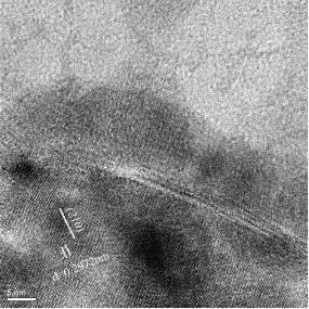 | Fig. 4 Laser alloyed dendrite structure by HRTEM for the area corresponding to area 2 in Fig. 1, upper part showing featureless except for cell structure |
 | Fig. 5 HRTEM micrograph, showing amorphous in featureless region and Al3Ni phase for dendrite. The inset is diffraction pattern corresponding to amorphous in featureless region. |
In Ni-Al laser alloyed layer, the melt was transformed in solidified structure that consists of dendrite and featureless region. The dendrite was relatively rich in Ni and was identified to be Al3Ni phase. Featureless region is high in Al that is surrounded by the dendrite, including less Ni, more Al and probable less P. Al3Ni and Al were not homogenously distributed in the laser alloyed region.
The authors have declared that no competing interests exist.
| [1] |
|
| [2] |
|
| [3] |
|
| [4] |
|
| [5] |
|
| [6] |
|
| [7] |
|
| [8] |
|
| [9] |
|
| [10] |
|
| [11] |
|
| [12] |
|
| [13] |
|